Gastric folds
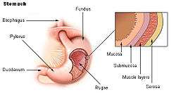
The gastric folds (or gastric rugae) are coiled sections of tissue that exist in the mucosal and submucosal layers of the stomach.[1] They provide elasticity by allowing the stomach to expand when a bolus enters it; these folds stretch outward through the action of mechanoreceptors which respond to the increase in pressure.[2] This allows the stomach to expand, therefore increasing the volume of the stomach without increasing pressure.[2] These folds provide the stomach with increased surface area for nutrient absorption during digestion.[2] Gastric folds may be seen during esophagogastroduodenoscopy or in radiological studies.[3][4]
| Gastric folds | |
|---|---|
 Stomach | |
| Details | |
| Identifiers | |
| Latin | plicae gastricae |
| TA | A05.5.01.028 |
| FMA | 75653 |
| Anatomical terminology | |
Layers
- Mucosa
This layer releases stomach acid. It is the innermost layer of the stomach[5] Affected by the hormone histamine which signals it to release Hydrocholoric acid (HCL).
- Sub-mucosa
This layer consists of different vessels and nerves, ganglion neurons, and adipose tissue. It is the second layer of the stomach and supports the mucosa.[6]
Gastric fold thickening
Thickening of the gastric folds may be observed by endoscopy or radiography and may aid in the differential diagnosis of many disease processes including:[3]
The folds become very thick due to inflammation.[7]
Ulcers cause breaks in the mucosa and cause erosion of the submucosa.
Gastrin levels increase due to tumors, which cause an increase in the gastric fold size.[7]
The mucosa pits are in excess causing thickening of the folds.[7]
- Carcinoma
- Helicobacter pylori infection
Causes inflammation of the folds.
- Gastric Syphilis[8]
- Cytomegalovirus
Mucosa change shape causing rugae enlargement.[9]
- Sarcoidosis
Causes thickening of the folds.
References
- David., Shier (2009). Hole's essentials of human anatomy & physiology. Butler, Jackie., Lewis, Ricki. (10th ed.). Boston: McGraw-Hill Higher Education. p. 421. ISBN 978-0077221355. OCLC 171614173.
- Michelle., McGuire (2013). Nutritional sciences : from fundamentals to food. Beerman, Kathy A. (3rd ed.). Belmont, CA: Wadsworth, Cengage Learning. p. 90. ISBN 978-0840058201. OCLC 786272310.
- L., Eisenberg, Ronald (2003). Gastrointestinal radiology : a pattern approach (4th ed.). Philadelphia: Lippincott Williams & Wilkins. pp. 223–236. ISBN 978-0781737067. OCLC 49550593.
- "The Stomach and Its Role in Digestion | Laparoscopic.MD". www.laparoscopic.md. Retrieved 14 November 2017.
- Taylor, Tim. "Stomach". InnerBody. Retrieved November 13, 2017.
- "Stomach". BioNet. Retrieved November 13, 2017.
- "Gastritis, Giant Hypertrophic - NORD (National Organization for Rare Disorders)". NORD (National Organization for Rare Disorders). Retrieved 9 December 2017.
- Butz, William; Watts, John; Rosales-Qiuntana, Sergio; Hicklin, Martin. "Erosive Gastritis as a Manifestation of Secondary Syphilis" (PDF). Retrieved 9 December 2017.
- Dughera, Francesca; Baino, Sara. "Cronkhite-Canada Syndrome". flipper.diff.org. Retrieved 9 December 2017.
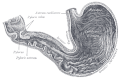 Drawing of the interior of the stomach.
| Authority control |
|---|