Cuneiform bones
There are three cuneiform ("wedge-shaped") bones in the human foot:
- the first or medial cuneiform
- the second or intermediate cuneiform, also known as the middle cuneiform
- the third or lateral cuneiform
| Cuneiform bones; Cuneiform | |
|---|---|
 Red=medial; yellow=intermediate; green=lateral | |
 Cuneiform bones of the left foot | |
| Details | |
| Identifiers | |
| Latin | os cuneiformis pl. ossa cuneiformia |
| FMA | 71339 |
| Anatomical terms of bone | |
They are located between the navicular bone and the first, second and third metatarsal bones and are medial to the cuboid bone.[1]
Structure
There are three cuneiform bones:
- The medial cuneiform (also known as first cuneiform) is the largest of the cuneiforms. It is situated at the medial side of the foot, anterior to the navicular bone and posterior to the base of the first metatarsal. Lateral to it is the intermediate cuneiform. It articulates with four bones: the navicular, second cuneiform, and first and second metatarsals. The tibialis anterior and fibularis longus muscle inserts at the medial cuneiform bone.[2]
- The intermediate cuneiform (second cuneiform or middle cuneiform) is shaped like a wedge, the thin end pointing downwards. The intermediate cuneiform is situated between the other two cuneiform bones (the medial and lateral cuneiforms), and articulates with the navicular posteriorly, the second metatarsal anteriorly and with the other cuneiforms on either side.
- The lateral cuneiform (also known as third cuneiform or external cuneiform) intermediate in size between the other two cuneiform bones, is also wedge-shaped, the base being uppermost. It occupies the center of the front row of the tarsal bones, between the intermediate cuneiform medially, the cuboid laterally, the navicular posteriorly and the third metatarsal in front. The tibialis posterior inserts at the medial cuneiform, while the flexor hallucis brevis originates from it.[2]
Muscle attachments
| Muscle | Direction | Attachment[2] |
| Tibialis anterior | Insertion | Medial cuneiform |
| Fibularis longus | Insertion | Medial cuneiform |
| Tibialis posterior | Insertion | Medial cuneiform |
| Flexor hallucis brevis | Origin | Lateral cuneiform |
Additional images
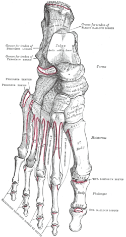 Bones of the right foot. Dorsal surface.
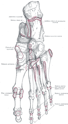 Bones of the right foot. Plantar Surface.
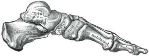 Skeleton of foot. Medial aspect.
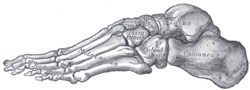 Skeleton of foot. Lateral aspect.
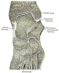 Oblique section of left intertarsal and tarsometatarsal articulations, showing the synovial cavities.
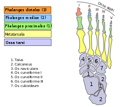 Bones of foot
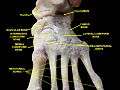 Cuneiform. Superior view.
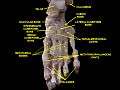 Cuneiform. Superior view.
Other animals
See also
- Cuneiform, for writing by pressing a wedge-shaped reed into wet clay.
References
- Bojsen-Møller, Finn; Simonsen, Erik B.; Tranum-Jensen, Jørgen (2001). Bevægeapparatets anatomi [Anatomy of the Locomotive Apparatus] (in Danish) (12th ed.). p. 245. ISBN 978-87-628-0307-7.
- Bojsen-Møller, Finn; Simonsen, Erik B.; Tranum-Jensen, Jørgen (2001). Bevægeapparatets anatomi [Anatomy of the Locomotive Apparatus] (in Danish) (12th ed.). pp. 364–367. ISBN 978-87-628-0307-7.
| Wikimedia Commons has media related to Cuneiform bones. |
This article is issued from
Wikipedia.
The text is licensed under Creative
Commons - Attribution - Sharealike.
Additional terms may apply for the media files.