First metatarsal bone
The first metatarsal bone is the bone in the foot just behind the big toe. The first metatarsal bone is the shortest of the metatarsal bones and by far the thickest and strongest of them.[1]
| First metatarsal bone | |
|---|---|
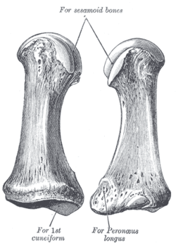 The first metatarsal. (Left.) | |
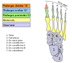 Bones of the right foot. Dorsal surface. The first metatarsal bone is shown in yellow farthest to the left | |
| Details | |
| Identifiers | |
| Latin | Os metatarsale I |
| Anatomical terms of bone | |
Like the four other metatarsals, it can be divided into three parts: base, body and head. The base is the part closest to the ankle and the head is closest to the big toe. The narrowed part in the middle is referred to as the body of the bone. The bone is somewhat flattened, giving it two sides: the plantar (towards the sole of the foot) and the dorsal side (the area facing upwards while standing).[1]
The base presents, as a rule, no articular facets (joint surfaces) on its sides, but occasionally on the lateral side there is an oval facet, by which it articulates with the second metatarsal. On the lateral part of the plantar surface there is a rough oval prominence, or tuberosity, for the insertion of the tendon of the fibularis longus.
The first metatarsal articulates (forms joints) with the medial cuneiform and to a small extent with the intermediate cuneiform bone.[2] Its proximal articular surface is large and kidney-shaped; its circumference is grooved, for the tarsometatarsal ligaments, and medially gives insertion to part of the tendon of the tibialis anterior.
The body of the bone is strong, and of well-marked prismoid form.
The head is large; on its plantar surface are two grooved facets on which the sesamoid bones glide; the facets are separated by a smooth elevation.
Muscle attachments
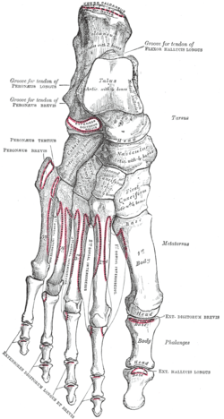 Muscle attachments (seen from above) | 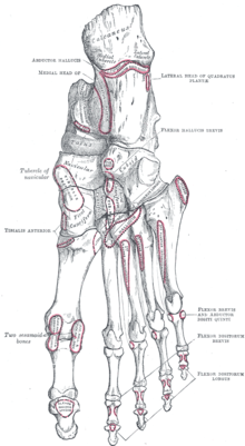 Muscle attachments (seen from below) |
Three muscles attach to the first metatarsal bone: the tibialis anterior, fibularis longus and first dorsal interosseus.[3]
The tibialis anterior inserts at the basis of the bone, while the fibularis longus inserts at the tuberosity. The lateral part of the first dorsal interosseus muscle originates from the medial side of the bone. Its function is to spread the toes.[4]
| Muscle | Direction | Attachment[3] |
|---|---|---|
| Tibialis anterior | Insertion | Basis of first metatarsal |
| Fibularis longus | Insertion | Tuberosity of first metatarsal |
| Dorsal interossei I | Origin | Lateral part of first metatarsal |
Additional images
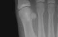 Sesamoid bones at the distal end of the first metatarsal.
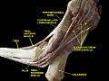 First metatarsal bone. Deep dissection.
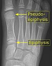 It is common in children to have a pseudo-epiphysis of the first metatarsal.[5]
References
This article incorporates text in the public domain from page 272 of the 20th edition of Gray's Anatomy (1918)
- Bojsen-Møller, Finn; Simonsen, Erik B.; Tranum-Jensen, Jørgen (2001). Bevægeapparatets anatomi [Anatomy of the Locomotive Apparatus] (in Danish) (12th ed.). p. 246. ISBN 978-87-628-0307-7.
- Platzer 2004, p. 218
- Bojsen-Møller, Finn; Simonsen, Erik B.; Tranum-Jensen, Jørgen (2001). Bevægeapparatets anatomi [Anatomy of the Locomotive Apparatus] (in Danish) (12th ed.). pp. 364–67. ISBN 978-87-628-0307-7.
- Bojsen-Møller, Finn; Simonsen, Erik B.; Tranum-Jensen, Jørgen (2001). Bevægeapparatets anatomi [Anatomy of the Locomotive Apparatus] (in Danish) (12th ed.). pp. 300–01. ISBN 978-87-628-0307-7.
- Mathis, SK; Frame, BA; Smith, CE (1989). "Distal first metatarsal epiphysis. A common pediatric variant". Journal of the American Podiatric Medical Association. 79 (8): 375–379. doi:10.7547/87507315-79-8-375. ISSN 8750-7315.
| Wikimedia Commons has media related to First metatarsal bone. |