Calcaneus
In humans, the calcaneus (/kælˈkeɪniəs/; from the Latin calcaneus or calcaneum, meaning heel[1]) or heel bone is a bone of the tarsus of the foot which constitutes the heel. In some other animals, it is the point of the hock.
| Calcaneus | |
|---|---|
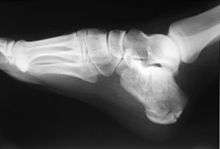
 The calcaneus forms the bony part of the heel. It forms a joint with the talus bone, the subtalar joint. | |
 Bones of the foot, with the calcaneus shown in red | |
| Details | |
| Identifiers | |
| Latin | Calcaneus, Calcaneum, Os calcis |
| MeSH | D002111 |
| TA | A02.5.11.001 |
| FMA | 24496 |
| Anatomical terms of bone | |
Structure
In humans, the calcaneus is the largest of the tarsal bones and the largest bone of the foot. The talus bone, calcaneus, and navicular bone are considered the proximal row of tarsal bones.[2] In the calcaneus, several important structures can be distinguished:[2]
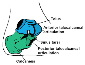
The half of the bone closest to the heel is the calcaneal tuberosity. On its lower edge on either side are its lateral and medial processes (serving as the origins of the abductor hallucis and abductor digiti minimi). The Achilles tendon is inserted into a roughened area on its superior side, the cuboid bone articulates with its anterior side, and on its superior side are three articular surfaces for the articulation with the talus bone. Between these superior articulations and the equivalents on the talus is the tarsal sinus (a canal occupied by the interosseous talocalcaneal ligament). At the upper and forepart of the medial surface of the calcaneus, below the middle talar facet, there is a horizontal eminence, the talar shelf (also sustentaculum tali), which gives attachment to the plantar calcaneonavicular (spring) ligament, tibiocalcaneal ligament, and medial talocalcaneal ligament. This eminence is concave above, and articulates with the middle calcaneal articular surface of the talus; below, it is grooved for the tendon of the flexor hallucis longus; its anterior margin gives attachment to the plantar calcaneonavicular ligament, and its medial margin to a part of the deltoid ligament of the ankle-joint.
On the lateral side is commonly a tubercle called the calcaneal tubercle (or trochlear process). This is a raised projection located between the tendons of the peroneus longus and brevis. It separates the two oblique grooves of the lateral surface of the calcaneus (for the tendons of the peroneal muscles).
Its chief anatomical significance is as a point of divergence of the previously common pathway shared by the distal tendons of peroneus longus and peroneus brevis en route to their distinct respective attachment sites. [2]
The calcaneus is part of two joints: the proximal intertarsal joint and the talocalcaneal joint. The point of the calcaneus is covered by the calcanean bursa.
Development
In the calcaneus, an ossification center, is developed during the 4th–7th week of fetal development. [2]
Function
Three muscles insert on the calcaneus: the gastrocnemius, soleus, and plantaris. These muscles are part of the posterior compartment of the leg and aid in walking, running and jumping. Their specific functions include plantarflexion of the foot, flexion of the knee, and steadying the leg on the ankle during standing. The calcaneus also serves as origin for several short muscles that run along the sole of the foot and control the toes.
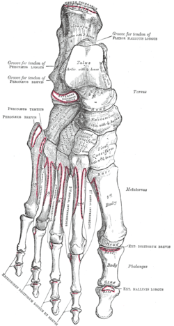 Muscle attachments (seen from above) | 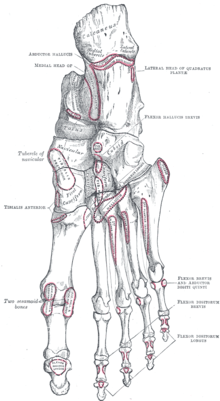 Muscle attachments (seen from below) |
| Muscle | Direction | Attachment[3] |
| Gastrocnemius | Insertion | Calcaneal tubercle through the achilles tendon |
| Soleus | Insertion | Calcaneal tubercle through the achilles tendon |
| Plantaris | Insertion | Calcaneal tubercle either directly or through the achilles tendon |
| Extensor digitorum brevis | Origin | Dorsal side of calcaneus |
| Abductor hallucis | Origin | Medial process of calcaneus |
| Extensor hallucis brevis | Origin | Dorsal side of calcaneus |
| Abductor digiti minimi | Origin | Calcaneal tubercle |
| Flexor digitorum brevis | Origin | Calcaneal tubercle |
| Quadratus plantae | Origin | Lateral and medial processes of calcaneus |
Clinical significance
Normally the tibia sits vertically above the calcaneus (pes rectus). If the calcaneal axis between these two bones is turned medially the foot is in an everted position (pes valgus), and if it is turned laterally the foot is in an inverted position (pes varus).[4]
- Calcaneal fracture, also known as Lover's fracture and Don Juan fracture
Disease
The talar shelf is typically involved in subtalar or talocalcaneal tarsal coalition.
See also
Additional images
 Left calcaneus. Animation.
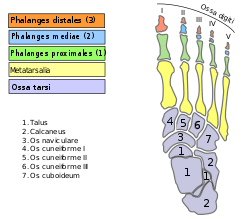 Bones of foot
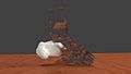 3D rendering of a left calcaneus derived from CT scan data. The calcaneus is white, and the other bones of the foot and ankle are clear to illustrate the position and relationship of the calcaneus to the other tarsal bones.
Notes
- Mosby’s Medical, Nursing and Allied Health Dictionary, Fourth Edition, Mosby-Year Book Inc., 1994, p. 242
- Platzer (2004), p 216
- Bojsen-Møller, Finn; Simonsen, Erik B.; Tranum-Jensen, Jørgen (2001). Bevægeapparatets anatomi [Anatomy of the Locomotive Apparatus] (in Danish) (12th ed.). pp. 364–367. ISBN 978-87-628-0307-7.
- Thieme Atlas of Anatomy (2006), p 410
References
- Platzer, Werner (2004). Color Atlas of Human Anatomy, Vol. 1: Locomotor System (5th ed.). Thieme. ISBN 3-13-533305-1.
- Thieme Atlas of Anatomy: General Anatomy and Musculoskeletal System. Thieme. 2006. ISBN 1-58890-419-9.
- Saladin, Kenneth (2012). Anatomy and Physiology, The Unity of Form and Function. McGraw Hill. pp. 270–271. ISBN 978-0-07-337825-1.
- "Calcaneus (Heel Bone) Fractures". American Academy of Orthopaedic Surgeons. Retrieved 13 Dec 2012.
External links
| Wikimedia Commons has media related to Calcaneus. |
- lljoints at The Anatomy Lesson by Wesley Norman (Georgetown University) (posterioranklejoint)
- 3D printable calcaneus model, free download in STL format (Embodi3D.com)
| Authority control |
|
|---|