Cuneiform cartilages
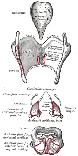
In the human larynx, the cuneiform cartilages (from Latin: cunei, "wedge-shaped"; also known as cartilages of Wrisberg) are two small, elongated pieces of yellow elastic cartilage, placed one on either side, in the aryepiglottic fold.[1]
| Cuneiform cartilages | |
|---|---|
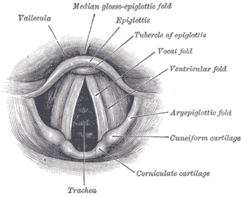 Laryngoscopic view of interior of larynx. | |
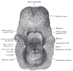 The entrance to the larynx, viewed from behind. | |
| Details | |
| Identifiers | |
| Latin | cartilagines cuneiformes |
| TA | A06.2.06.001 |
| FMA | 55111 |
| Anatomical terminology | |
The cuneiforms are paired cartilages that sit on top of and move with the arytenoids.[2] They are located above and in front of the corniculate cartilages, and the presence of these two pairs of cartilages result in small bulges on the surface of the mucous membrane.[3] Covered by the aryepiglottic folds, the cuneiforms form the lateral aspect of the laryngeal inlet, while the corniculates form the posterior aspect, and the epiglottis the anterior.[4]
Function of the cuneiform cartilages is to support the vocal folds and lateral aspects of the epiglottis. They also provide a degree of solidity to the folds in which they are embedded.[3]
Additional images
 Posterior view of the cartilages of the larynx
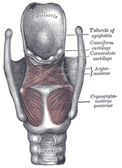 Posterior view of the muscles of the larynx
References
This article incorporates text in the public domain from page 1075 of the 20th edition of Gray's Anatomy (1918)
- Gray's Anatomy (1918), see infobox
- Rosen, Clarke A.; Simpson, Blake (2008). Operative Techniques in Laryngology. Springer. Retrieved November 2013. Check date values in:
|accessdate=(help) - Seikel, J. Anthony; King, Douglas W.; Drumright, David G. (2010). Anatomy & Physiology for Speech, Language, and Hearing (4th ed.). Delmar, NY: Cengage Learning. ISBN 978-1-4283-1223-4.
- The Essentials of Respiratory Care. Elsevier Health Sciences. 2005-01-07. p. 77. ISBN 9780323027007. Retrieved November 2013. Check date values in:
|accessdate=(help)
| Authority control |
|---|