Alpha wave
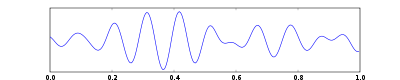
Alpha waves are neural oscillations in the frequency range of 8–12 Hz[1] arising from the synchronous and coherent (in phase or constructive) electrical activity of thalamic pacemaker cells in humans. They are also called Berger's waves after the founder of EEG.
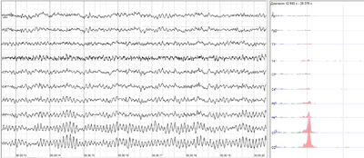
Alpha waves are one type of brain waves detected either by electroencephalography (EEG) or magnetoencephalography (MEG), and can be quantified using quantitative electroencephalography (qEEG). They predominantly originate from the occipital lobe during wakeful relaxation with closed eyes. Alpha waves are reduced with open eyes, drowsiness and sleep. Historically, they were thought to represent the activity of the visual cortex in an idle state. More recent papers have argued that they inhibit areas of the cortex not in use, or alternatively that they play an active role in network coordination and communication.[2] Occipital alpha waves during periods of eyes closed are the strongest EEG brain signals.
An alpha-like variant called a mu wave can be found over the primary motor cortex.
History of alpha waves
Alpha waves were discovered by German neurologist Hans Berger, the inventor of the EEG itself. Alpha waves were among the first waves documented by Berger, along with beta waves, and he displayed an interest in "alpha blockage", the process by which alpha waves decrease and beta waves increase upon a subject opening their eyes. This distinction earned the alpha wave the alternate title of "Berger's Wave".
Berger took a cue from Ukrainian physiologist Vladimir Pravdich-Neminsky, who used a string galvanometer to create a photograph of the electrical activity of a dog's brain. Using similar techniques, Berger confirmed the existence of electrical activity in the human brain. He first did this by presenting a stimulus to hospital patients with skull damage and measuring the electrical activity in their brains. Later he ceased the stimulus method and began measuring the natural rhythmic electrical cycles in the brain. The first natural rhythm he documented was what would become known as the alpha wave. Berger was very thorough and meticulous in his data-gathering, but despite his brilliance, he did not feel confident enough to publish his discoveries until at least five years after he had made them. In 1929, he published his first findings on alpha waves in the journal Archiv für Psychiatrie. He was originally met with derision for his EEG technique and his subsequent alpha and beta wave discoveries. His technique and findings did not gain widespread acceptance in the psychological community until 1937, when he gained the approval of the famous physiologist Lord Adrian, who took a particular interest in alpha waves.[3]
Alpha waves again gained recognition in the early 1960s and 1970s with the creation of a biofeedback theory relating to brain waves (see below). Such biofeedback, referred to as a kind of neurofeedback, relating to alpha waves is the conscious elicitation of alpha brainwaves by a subject. Two researchers in the United States explored this concept through unrelated experiments. Joe Kamiya, of the University of Chicago, discovered that some individuals had the conscious ability to recognize when they were creating alpha waves, and could increase their alpha activity. These individuals were motivated through a reward system from Kamiya. The second progenitor of biofeedback is Barry Sterman, from the University of California, Los Angeles. He was working with monitoring brain waves in cats and found that, when the cats were trained to withhold motor movement, they released SMR, or mu, waves, a wave similar to alpha waves. Using a reward system, he further trained these cats to enter this state more easily. Later, he was approached by the United States Air Force to test the effects of a jet fuel that was known to cause seizures in humans. Sterman tested the effects of this fuel on the previously-trained cats, and discovered that they had a higher resistance to seizures than non-trained cats.
Alpha wave biofeedback has gained interest for having some successes in humans for seizure suppression and for treatment of depression.[4]
Alpha waves again gained interest in regards to an engineering approach to the science fiction challenge of psychokinesis, i.e. control of movement of a physical object using energy emanating from a human brain. In 1988, EEG alpha rhythm was used in a Brain-Computer Interface experiment of control of a movement of a physical object, a robot. [5][6]It was the first experiment to demonstrate control of a physical object, a robot, using EEG. [7][8]
Types of alpha waves
Some researchers posit that there are at least two forms of alpha waves, which may have different functions in the wake-sleep cycle.
Alpha waves are present at different stages of the wake-sleep cycle. The most widely researched is during the relaxed mental state, where the subject is at rest with eyes closed, but is not tired or asleep. This alpha activity is centered in the occipital lobe, and is presumed to originate there, although there has been recent speculation that it instead has a thalamic origin.[9] This wave begins appearing at around four months, and is initially a frequency of 4 waves per second. The mature alpha wave, at 10 waves per second, is firmly established by age 3.[10]
The second occurrence of alpha wave activity is during REM sleep. As opposed to the awake form of alpha activity, this form is located in a frontal-central location in the brain. The purpose of alpha activity during REM sleep has yet to be fully understood. Currently, there are arguments that alpha patterns are a normal part of REM sleep, and for the notion that it indicates a semi-arousal period. It has been suggested that this alpha activity is inversely related to REM sleep pressure.
It has long been believed that alpha waves indicate a wakeful period during sleep. This has been attributed to studies where subjects report non-refreshing sleep and have EEG records reporting high levels of alpha intrusion into sleep. This occurrence is known as alpha wave intrusion.[11] However, it is possible that these explanations may be misleading, as they only focus on alpha waves being generated from the occipital lobe.
Alpha wave intrusion
Alpha wave intrusion occurs when alpha waves appear with non-REM sleep when delta activity is expected. It is hypothesized to be associated with fibromyalgia,[12] although the study may be inadequate due to a small sampling size.
Despite this, alpha wave intrusion has not been significantly linked to any major sleep disorder, including fibromyalgia, chronic fatigue syndrome, and major depression. However, it is common in chronic fatigued patients, and may amplify the effects of other sleep disorders.[13]
Mistake prediction
Following this lapse-of-attention line of thought, a recent study indicates that alpha waves may be used to predict mistakes. In it, MEGs measured increases of up to 25% in alpha brain wave activity before mistakes occurred. This study used common sense: alpha waves indicate idleness, and mistakes are often made when a person is doing something automatically, or "on auto-pilot", and not paying attention to the task they are performing. After the mistake was noticed by the subject, there was a decrease in alpha waves as the subject began paying more attention. This study hopes to promote the use of wireless EEG technology on employees in high-risk fields, such as air traffic controlling, to monitor alpha wave activity and gauge the attention level of the employee.[14]
Alpha wave artifacts
As demonstrated by Dr. Adrian R. M. Upton[15], it is possible for extraneous sources (ambient fluctuations detected with a mound of Jell-O in Upton's experiments) to cause signals to appear on an EEG readout, causing false signals to be interpreted as healthy alpha waves. This finding suggests that it is possible that a non-flat EEG could lead to the interpretation that a patient is still living when in fact he or she is long dead.
Cecil Adams from The Straight Dope discusses this scenario:
Sometimes it's claimed Jell-O brainwaves are identical to a healthy adult's. That's clearly a stretch, but the Jell-O EEG readings do look pretty similar to a normal human alpha rhythm. Alpha waves are observed when a patient is awake and resting with eyes closed, and in some kinds of sleep and reversible coma. True, the Jell-O waves are a little slower and of much lower amplitude, barely within normal human limits, but that doesn't tell you much by itself. Hypoxia, encephalitis, and other medical conditions can cause reduced frequency and amplitude, as can drug use.[16]
See also
Brain waves
- Delta wave – (0.5 – 3 Hz)
- Theta wave – (4 – 7 Hz)
- Alpha wave – (7 – 15 Hz)
- Mu wave – (7.5 – 12.5 Hz)
- SMR wave – (12.5 – 15.5 Hz)
- Beta wave – (15 – 30 Hz)
- Gamma wave – (>30 Hz)
References
- Foster, JJ; Sutterer, DW; Serences, JT; Vogel, EK; Awh, E (July 2017). "Alpha-Band Oscillations Enable Spatially and Temporally Resolved Tracking of Covert Spatial Attention". Psychological Science. 28 (7): 929–941. doi:10.1177/0956797617699167. PMC 5675530. PMID 28537480.
- Palva S.; Palva J.M. (2007). "New vistas for a-frequency band oscillations". Trends Neurosci. 30 (4): 150–158. doi:10.1016/j.tins.2007.02.001. PMID 17307258.
- Karbowski K (2002). "Hans Berger (1873-194)". Journal of Neurology. 249 (8): 1130–1131. doi:10.1007/s00415-002-0872-4. PMID 12420722.
- Ulrich Kraft. Train Your Brain-Mental exercises with neurofeedback may ease symptoms of attention-deficit disorder, epilepsy and depression--and even boost cognition in healthy brains. Scientific American. 2006
- S. Bozinovski, M. Sestakov, L. Bozinovska: Using EEG alpha rhythm to control a mobile robot, In G. Harris, C. Walker (eds.) Proc. IEEE Annual Conference of Medical and Biological Society, p. 1515-1516, New Orleans, 1988
- S. Bozinovski: Mobile robot trajectory control: From fixed rails to direct bioelectric control, In O. Kaynak (ed.) Proc. IEEE Workshop on Intelligent Motion Control, p. 63-67, Istanbul, 1990
- M. Lebedev: Augmentation of sensorimotor functions with neural prostheses. Opera Medica and Physiologica. Vol. 2 (3): 211-227, 2016
- M. Lebedev, M. Nicolelis: Brain-machine interfaces: from basic science to neuroprostheses and neurorehabilitation, Physiological Review 97:737-867, 2017
- Domino E. F.; Ni L. S.; et al. (2009). "Tobacco smoking produces widespread dominant brainwave alpha frequency increases". International Journal of Psychophysiology. 74 (3): 192–198. doi:10.1016/j.ijpsycho.2009.08.011. PMC 2788071. PMID 19765621.
- Niedermeyer E (1997). "Alpha rhythms as physiological and abnormal phenomena". International Journal of Psychophysiology. 26 (1–3): 31–49. doi:10.1016/s0167-8760(97)00754-x. PMID 9202993.
- Task Force Allas (1992). "ASDA report on EEG arousals: scoring rules and examples". Sleep. 15 (2): 173–184. doi:10.1093/sleep/15.2.173.
- Germanowicz D, Lumertz MS, Martinez D, Margarites AF (2006). "Sleep disordered breathing concomitant with fibromyalgia syndrome". J Bras Pneumol. 32 (4): 333–8. doi:10.1590/s1806-37132006001100012. PMID 17268733.
- Manu, Peter; Lane, Thomas J.; Matthews, Dale A.; Castriotta, Richard J.; Watson, Robert K.; Abeles, Micha (1994). "Alpha-delta sleep in patients with a chief complaint of chronic fatigue". Southern Medical Journal. 87 (4): 465–470. doi:10.1097/00007611-199404000-00008.
- "Brain Wave Patterns Can Predict Blunders, New Study Finds". UC Davis News and Information. University of California, Davis campus. 23 March 2009.
- JELL‐0 TEST FINDS LIFELIKE SIGNAL, By BOYCE RENSBERGER, New York Times, MARCH 6, 1976
- "Can brainwaves be detected in lime Jell-O?". straightdope.com. 11 June 2010. Retrieved 7 April 2018.
Further reading
- Brazier, M. A. B. (1970), The Electrical Activity of the Nervous System, London: Pitman