


Retortamonas intestinalis
 ShareCompartir
[Retortamonas intestinalis]
Causal Agents
Retortamonas intestinalis, a nonpathogenic flagellate.
Life Cycle
Both cysts and trophozoites of Retortamonas intestinalis are shed in feces . Infection occurs after the ingestion of cysts in fecal-contaminated food or water, or on fomites
 . In the large (and possibly small) intestine, excystation releases trophozoites. Retortamonas resides in the large intestine, where it is regarded as a commensal and is not known to cause disease.
Geographic Distribution
Worldwide.
Clinical Presentation
Retortamonas intestinalis is considered nonpathogenic. The presence of trophozoites and/or cysts in stool specimens can however be an indicator of fecal contamination of a food or water source, and thus does not rule-out other parasitic infections.
Retortamonas intestinalis, trophozoites.
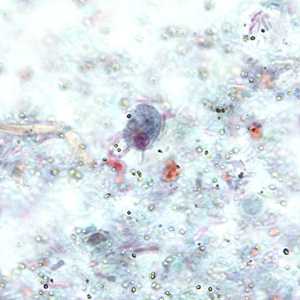
Figure A: Trophozoite of R. intestinalis in a stool specimen, stained with trichrome.
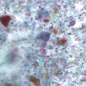
Figure B: Trophozoite of R. intestinalis in a stool specimen, stained with trichrome.
Retortamonas intestinalis, cysts.
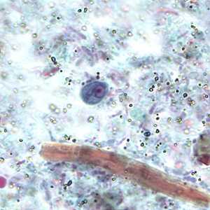
Figure A: Cyst of R. intestinalis in a stool specimen, stained with trichrome.
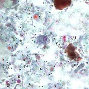
Figure B: Cyst of R. intestinalis in a stool specimen, stained with trichrome.
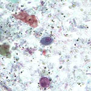
Figure C: Cyst of R. intestinalis in a stool specimen, stained with trichrome.
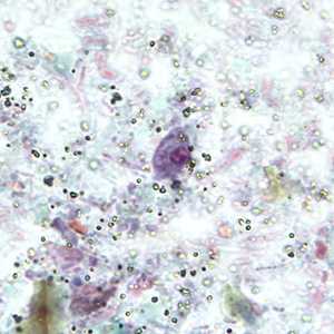
Figure D: Cyst of R. intestinalis in a stool specimen, stained with trichrome.
Laboratory Diagnosis
Retortamonas intestinalis is identified through the detection of trophozoites and/or cysts in stool specimens. Identification is best accomplished by direct wet mounts that reveal the characteristic, jerky movement of the organisms. They may also be identified in permanent stained smears, although their affinities for stain are inconsistent and with their small size are often overlooked.
Treatment Information
As this species is considered nonpathogenic, there are no treatment recommendations for this organism.
DPDx is an education resource designed for health professionals and laboratory scientists. For an overview including prevention and control visit www.cdc.gov/parasites/.
- Page last reviewed: May 3, 2016
- Page last updated: May 3, 2016
- Content source:
- Global Health – Division of Parasitic Diseases and Malaria
- Notice: Linking to a non-federal site does not constitute an endorsement by HHS, CDC or any of its employees of the sponsors or the information and products presented on the site.
- Maintained By: