


Case #448 - July, 2017
 ShareCompartir
A lung biopsy was obtained on a 29-year-old-patient from Cameroon with cough, dyspnea, and hemoptysis. Figures A–D show what was observed on a hematoxylin and eosin (H & E) stained section; Figure A is at 200x, Figure B 400x, and Figures C and D at 1000x magnification. What is your diagnosis? Based on what criteria?
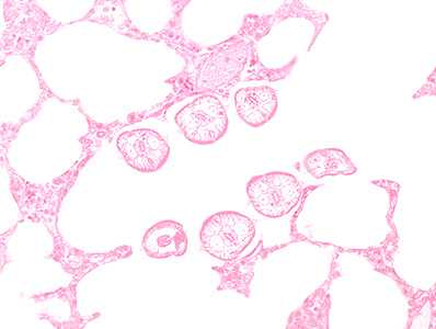
Figure A
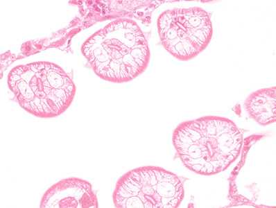
Figure B
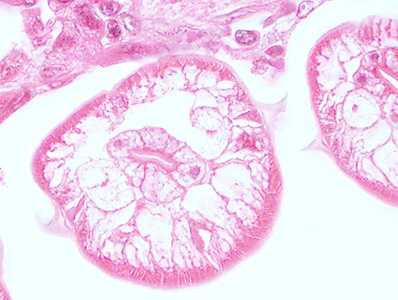
Figure C
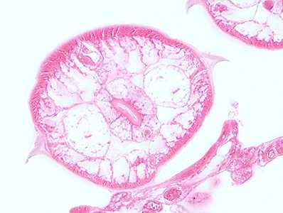
Figure D
Case Answer
This was a case of ascariasis based on the presence of migrating larvae. Diagnostic morphologic features shown in the images included:
- Prominent alae.
- Large excretory canals.
- Polymyarian muscle structure.
- Intestine lined with microvilli.
For more information on: ascariasis
Images presented in the monthly case studies are from specimens submitted for diagnosis or archiving. On rare occasions, clinical histories given may be partly fictitious.
DPDx is an education resource designed for health professionals and laboratory scientists. For an overview including prevention and control visit www.cdc.gov/parasites/.
- Page last reviewed: September 19, 2017
- Page last updated: September 20, 2017
- Content source:
- Global Health – Division of Parasitic Diseases and Malaria
- Notice: Linking to a non-federal site does not constitute an endorsement by HHS, CDC or any of its employees of the sponsors or the information and products presented on the site.
- Maintained By: