


Case #443 - May, 2017
 ShareCompartir
A 7-year-old female who had recently moved from the Democratic Republic of Congo to the US was treated for mild, non-immune hemolytic anemia and appeared to be in stable condition. Blood smears were made and stained with Wright-Giemsa at the time of her evaluation. Figures A – K show what was observed. What is your diagnosis? Based on what criteria? What additional tests, if any, should be performed for this case?
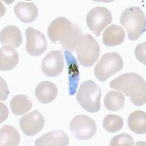
Figure A
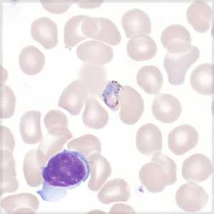
Figure B
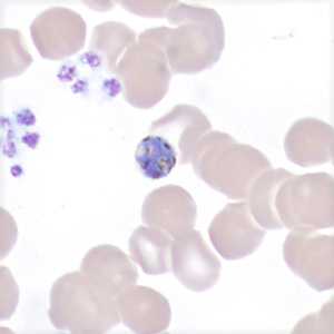
Figure C
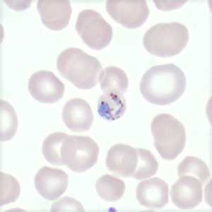
Figure D
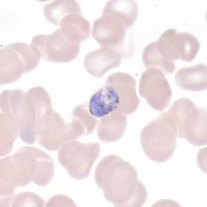
Figure E
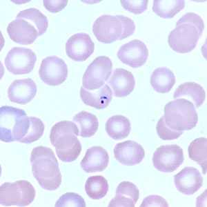
Figure F
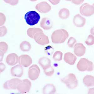
Figure G
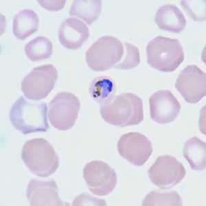
Figure H
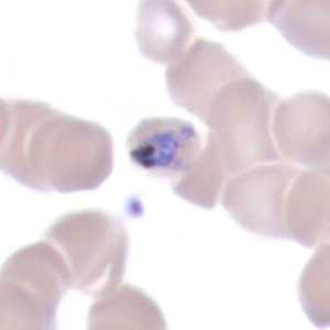
Figure I
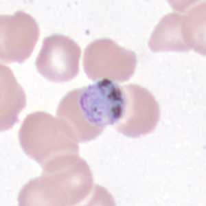
Figure J
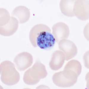
Figure K
Case Answer
This was a case of mixed infection of malaria caused by P. falciparum, P. malariae and P. ovale (confirmed by PCR). Diagnostic features shown were:
- Reduced, slightly enlarged as well as normal sized infected red blood cells.
- P falciparum gametocyte (Figure A), ring (Figure F).
- P. malariae trophozoites, “basket forms” (Figures B, D).
- P. malariae trophozoites, “band form” (Figure I).
More on Plasmodium species: Malaria
Images presented in the monthly case studies are from specimens submitted for diagnosis or archiving. On rare occasions, clinical histories given may be partly fictitious.
DPDx is an education resource designed for health professionals and laboratory scientists. For an overview including prevention and control visit www.cdc.gov/parasites/.
- Page last reviewed: July 19, 2017
- Page last updated: July 19, 2017
- Content source:
- Global Health – Division of Parasitic Diseases and Malaria
- Notice: Linking to a non-federal site does not constitute an endorsement by HHS, CDC or any of its employees of the sponsors or the information and products presented on the site.
- Maintained By: