


Case #439 - March, 2017
 ShareCompartir
A 29-year-old male immigrant from Nigeria presented to an emergency room with fever, splenomegaly, jaundice, and neurologic changes. Blood was drawn and used for a complete blood count (CBC) and complete blood chemistries panel which revealed anemia, thrombocytopenia, hypoglycemia, mildly elevated bilirubin and lactic acidosis. Figures A and B show what was observed on a Wright-Giemsa stained thin blood smear. What is your diagnosis? Based on what criteria?
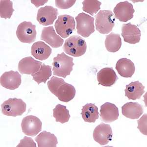
Figure A
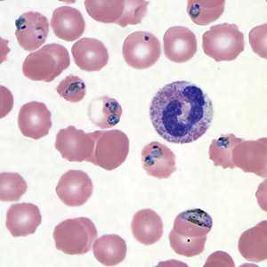
Figure B
Case Answer
This was a case of severe malaria caused by Plasmodium falciparum. Diagnostic features shown were:
- normal sized infected red blood cells
- parasite density estimated to be greater than 5% of red blood cells. Note that virtually all of the parasites present as compact trophozoites with pigment.
- multiply infected red blood cells.
- accolade/appliqué
- an immature P. falciparum gametocyte (Figure B), also in a normal sized red blood cell.
This case demonstrates the abnormal morphology of parasites observed when blood smears for the diagnosis of malaria are not made in a timely manner. These parasites have continued to develop outside of the body within the collected blood tube prior to the diagnostic smears being made. Other observations included both infected and non-infected red blood cells showing crenation.
For more on malaria:
Images presented in the monthly case studies are from specimens submitted for diagnosis or archiving. On rare occasions, clinical histories given may be partly fictitious.
DPDx is an education resource designed for health professionals and laboratory scientists. For an overview including prevention and control visit www.cdc.gov/parasites/.
- Page last reviewed: April 19, 2017
- Page last updated: April 19, 2017
- Content source:
- Global Health – Division of Parasitic Diseases and Malaria
- Notice: Linking to a non-federal site does not constitute an endorsement by HHS, CDC or any of its employees of the sponsors or the information and products presented on the site.
- Maintained By: