


Case #433 - December, 2016
 ShareCompartir
A 34-year-old male from Louisiana presented with abdominal discomfort. A stool specimen was collected in 10% formalin and a formalin-ethyl acetate (FEA) concentration was performed. A wet mount smear was prepared from the sediment and examined for ova and parasites (O & P). Figure A was captured at 100x magnification; Figures B-D were captured at 400x magnification. The objects of interest measured 155-165μm by 48-55μm. What is your diagnosis? Based on what criteria?
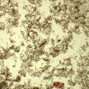
Figure A
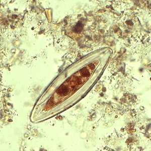
Figure B
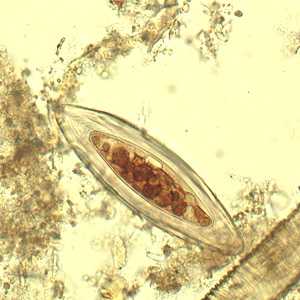
Figure C
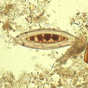
Figure D
Case Answer
The objects shown are artifacts commonly known as “Beaver bodies.” They are algae (Psorospermium haeckelli) that can be present in the tissues of crayfish and can be found on O & P examinations of individuals that recently consumed crayfish.
More on: artifacts
This case and images were kindly provided by ARUP Laboratories, Salt Lake City, UT
Images presented in the monthly case studies are from specimens submitted for diagnosis or archiving. On rare occasions, clinical histories given may be partly fictitious.
DPDx is an education resource designed for health professionals and laboratory scientists. For an overview including prevention and control visit www.cdc.gov/parasites/.
- Page last reviewed: January 10, 2017
- Page last updated: January 10, 2017
- Content source:
- Global Health – Division of Parasitic Diseases and Malaria
- Notice: Linking to a non-federal site does not constitute an endorsement by HHS, CDC or any of its employees of the sponsors or the information and products presented on the site.
- Maintained By: