


Case #424 - July 2016
 ShareCompartir
A 22-month-old female presented to the emergency room after 10 days of cyclic fever and chills, decreased PO intake, and seizures. The child had emigrated from Afghanistan with her family three months prior. Blood specimens were collected for routine work-up. A CBC was flagged for low platelet count, and smears were made and stained with Wright-Giemsa. During microscopic examination of thin smears, a technologist identified possible malaria parasites. Images were captured and sent to the DPDx Team for diagnostic assistance. Figures A-F show six of the images received for consultation. What is your diagnosis? Based on what criteria?
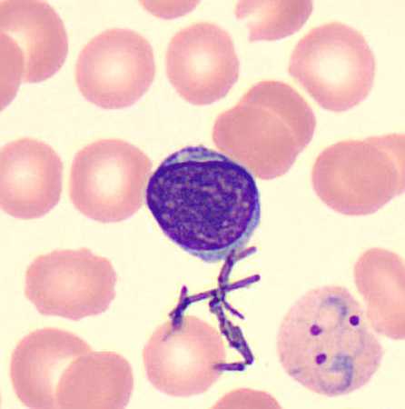
Figure A
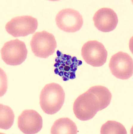
Figure B
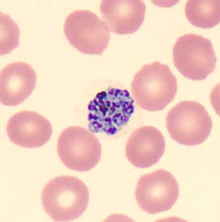
Figure C
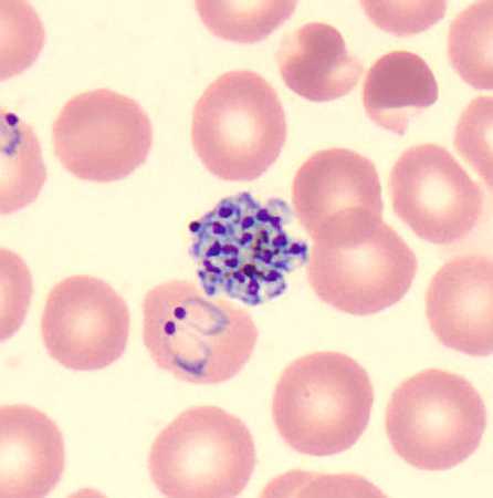
Figure D
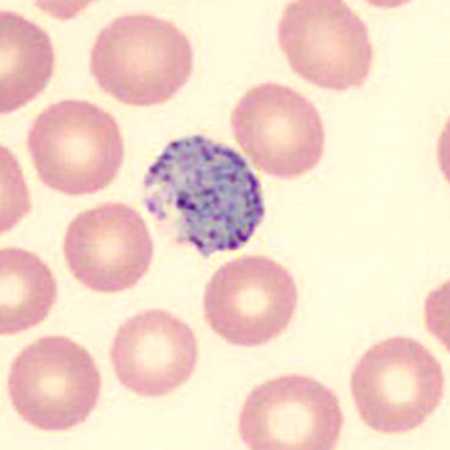
Figure E
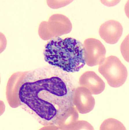
Figure F
Case Answer
This was a case of malaria caused by Plasmodium vivax. Diagnostic morphologic features included:
- sturdy rings with a large, single chromatin dot (Figures A and D).
- large round gametocytes with fine pigment (Figure E).
- schizonts with greater than 13 merozoites (Figures B-D); the object in Figure F shows a multiply-infected RBC that contains both a gametocyte and a schizont.
Figure A also showed bacterial contaminants. In addition to the morphologic features listed above, the patient’s travel history in Afghanistan was also supportive.
More on: Malaria
Acknowledgements
This case and images were kindly provided by Texas Children’s Hospital, Houston, TX.
DPDx is an education resource designed for health professionals and laboratory scientists. For an overview including prevention and control visit www.cdc.gov/parasites/.
- Page last reviewed: August 24, 2016
- Page last updated: August 24, 2016
- Content source:
- Global Health – Division of Parasitic Diseases and Malaria
- Notice: Linking to a non-federal site does not constitute an endorsement by HHS, CDC or any of its employees of the sponsors or the information and products presented on the site.
- Maintained By: