


Case #423 - July 2016
 ShareCompartir
A six-year-old boy from Washington, D.C. presented with a non-migratory furuncle on his scalp several weeks after returning from a trip to Belize (Figures A and B). A small incision was made and a worm-like organism (Figure C) was removed. The case was diagnosed as furuncular myiasis and the specimen was sent to the CDC-DPDx for identification. Figure D shows a close-up of the posterior spiracles. What is your identification? Based on what criteria?
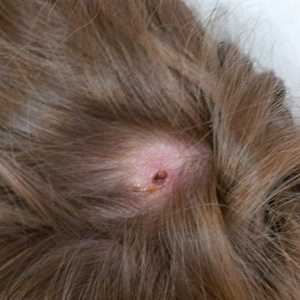
Figure A
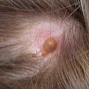
Figure B
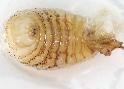
Figure C
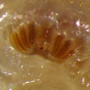
Figure D
Case Answer
This was a case of furuncular myiasis caused by Dermatobia hominis. Diagnostic morphologic features included:
- a robust larva with cuticular spines not present on the terminal three body segments (Figure C).
- spiracular plates lacking a peritreme and containing three slightly-curved slits (Figure D); the presence of three slits indicated this was a third-instar larva.
In addition to the morphologic features, the clinical presentation and travel history to Belize were also both supportive. Dermatobia hominis is endemic to the American tropics and is one of the most common causes of myiasis in people visiting Central and South America and the Caribbean.
More on: Myiasis
Acknowledgements
This case and images, in part, were kindly provided by Christie Athalia, Centers for Disease Control and Prevention, Washington, D.C.
DPDx is an education resource designed for health professionals and laboratory scientists. For an overview including prevention and control visit www.cdc.gov/parasites/.
- Page last reviewed: August 24, 2016
- Page last updated: August 24, 2016
- Content source:
- Global Health – Division of Parasitic Diseases and Malaria
- Notice: Linking to a non-federal site does not constitute an endorsement by HHS, CDC or any of its employees of the sponsors or the information and products presented on the site.
- Maintained By: