


Case #417 - April 2016
 ShareCompartir
A 27-year-old man had worms excised from his eyelid and chest at a health care facility in Canada. The patient had traveled to Sudan two years earlier, but had not traveled since then. The worms were sent to the DPDx Team for diagnostic assistance. The worm which was removed from the eyelid (Figure A) measured 55 mm in length. Figures B-D show the anterior, middle, and posterior portions of the same worm, respectively, at 100x magnification. A thin cross-section was made with a scalpel at approximately mid-body and examined. Figures E and F show what was observed at 100x and 200x magnification, respectively, of the cross section. What is your diagnosis? Based on what criteria?
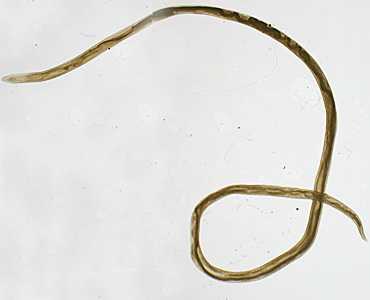
Figure A
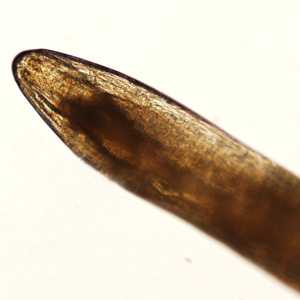
Figure B
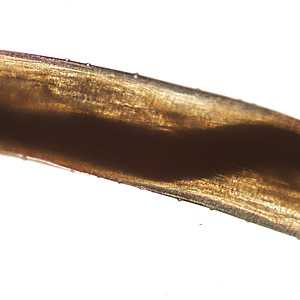
Figure C
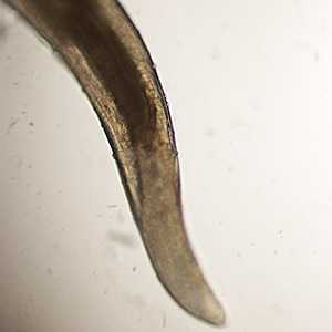
Figure D
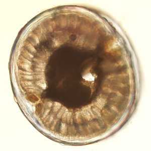
Figure E
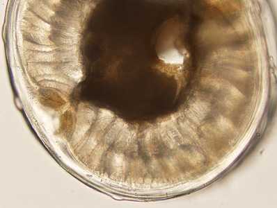
Figure F
Case Answer
This was a case of loiasis caused by the African eye worm, Loa loa. Morphologic features shown in the images included:
•presence of irregularly spaced bosses, best seen in Figure C (blue arrowheads).
•a moderately thick cuticle shown in Figures E and F (bosses can also be seen, blue arrowheads).
•8-10 coelomyarian muscle cells per quadrant (Figures E and F).
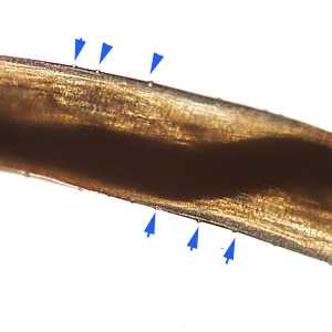
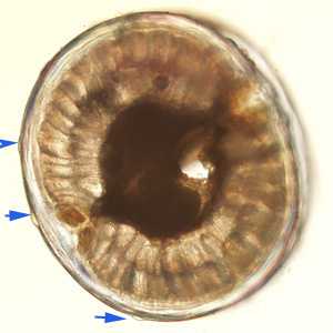
Figure E
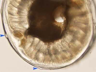
Figure F
More on: loiasis
Acknowledgements
This case was kindly provided by The Provincial Laboratory of Public Health, University of Alberta, Edmonton, Canada
DPDx is an education resource designed for health professionals and laboratory scientists. For an overview including prevention and control visit www.cdc.gov/parasites/.
- Page last reviewed: August 24, 2016
- Page last updated: August 24, 2016
- Content source:
- Global Health – Division of Parasitic Diseases and Malaria
- Notice: Linking to a non-federal site does not constitute an endorsement by HHS, CDC or any of its employees of the sponsors or the information and products presented on the site.
- Maintained By: