


Case #410 - December 2015
 ShareCompartir
A 22-year-old man, who had traveled to Kenya for a month, developed fever, chills, and general weakness within four days after returning to the U.S. He sought medical attention at a local hospital and admitted to not being fully compliant with taking his anti-malarial medication. Blood smears were ordered, stained with Wright-Giemsa stain, and examined at 1000x magnification with oil. Figures A-I show what was observed on a stained thin smear. What is your diagnosis? Based on what criteria?
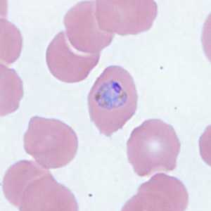
Figure A
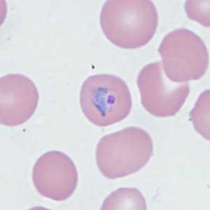
Figure B
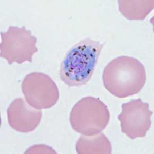
Figure C
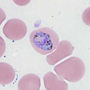
Figure D
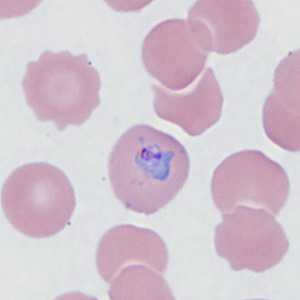
Figure E

Figure F
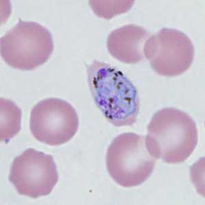
Figure G
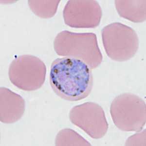
Figure H
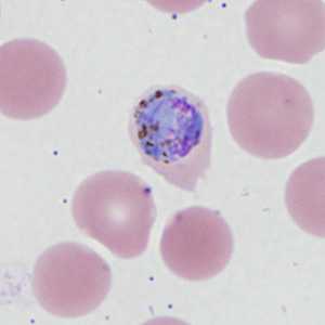
Figure I
Case Answer
This was a case of malaria caused by Plasmodium ovale. Diagnostic morphologic features shown included:
- sturdy, compact developing trophozoites in normal to slightly-enlarged RBCs (Figures A, B, and E).
- gametocytes with coarse pigment (Figures C, D, F, H, and I).
- an immature schizont with coarse pigment (Figure G).
- infected RBCs that demonstrated elongation and fimbriation (Figures A, C, F, G and I).
In addition to the morphologic features mentioned above, the travel to Kenya was also supportive. Schüffner’s stippling was not observed in any of the infected RBCs because the smears were stain with Wright-Giemsa stain. This case highlights the importance of using Giemsa stain, at a pH at or near 7.2, in helping to ensure an accurate identification is made.
More on: Malaria
DPDx is an education resource designed for health professionals and laboratory scientists. For an overview including prevention and control visit www.cdc.gov/parasites/.
- Page last reviewed: August 24, 2016
- Page last updated: August 24, 2016
- Content source:
- Global Health – Division of Parasitic Diseases and Malaria
- Notice: Linking to a non-federal site does not constitute an endorsement by HHS, CDC or any of its employees of the sponsors or the information and products presented on the site.
- Maintained By: