


Case #404 - September 2015
 ShareCompartir
A 31-year-old male presented with recurrent anal warts first noticed the summer of 2014. The warts were biopsied and diagnosed as low grade anal intraepithelial neoplasia (AIN1) in March 2015. Other symptoms included mild discomfort with defecation. In September 2015, an anal Pap smear was performed and stained with Papanicolau. Figures A-E show what was observed by the attending pathologist. The objects of interest ranged in size from 7-15 micrometers. What is your diagnosis? Based on what criteria?
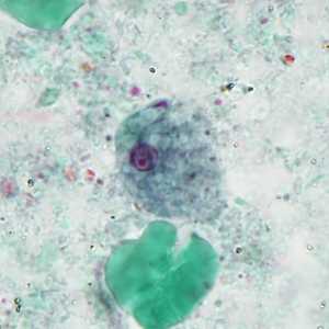
Figure A
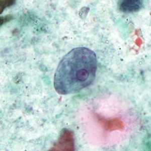
Figure B
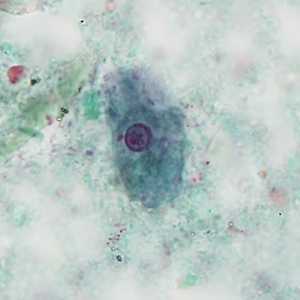
Figure C
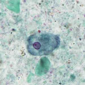
Figure D
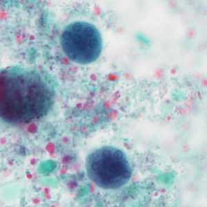
Figure E
Case Answer
The images in this case showed trophozoites and cysts of the nonpathogenic intestinal amoeba, Entamoeba coli. Diagnostic morphologic features included:
- trophozoites (Figures A-D) within the size range for E. coli with vacuolated cytoplasm and a single nucleus that contains irregular peripheral chromatin and an eccentric karyosome.
- mature cysts (Figure E) with more than four nuclei.
It is important to report nonpathogenic protozoa in all stool O&P examinations as their presence may be indicative of fecal contamination of a food or water source.
More on: Intestinal Amoebae
Images presented in the monthly case studies are from specimens submitted for diagnosis or archiving. On rare occasions, clinical histories given may be partly fictitious.
DPDx is an education resource designed for health professionals and laboratory scientists. For an overview including prevention and control visit www.cdc.gov/parasites/.
- Page last reviewed: August 24, 2016
- Page last updated: August 24, 2016
- Content source:
- Global Health – Division of Parasitic Diseases and Malaria
- Notice: Linking to a non-federal site does not constitute an endorsement by HHS, CDC or any of its employees of the sponsors or the information and products presented on the site.
- Maintained By: