


Case #401 - August 2015
 ShareCompartir
A worm approximately three centimeters long was observed and removed during a routine colonoscopy of a 54-year-old man from Scandinavia. The worm was sent to the CDC-DPDx Team for identification. Figure A shows the gross worm at 10x magnification using a dissecting microscope. It was then cleared with lacto-phenol and examined at 100x magnification (Figures B and C show the anterior and posterior respectively). A thin cross-section was made and examined at the same magnification (Figure D) and 200x (Figure E). What is your diagnosis? Based on what criteria?
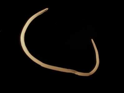
Figure A
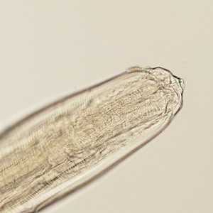
Figure B
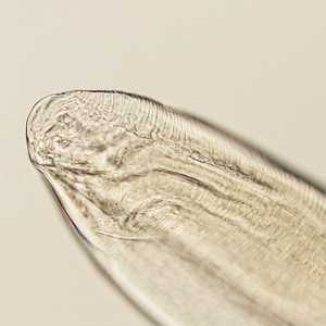
Figure C
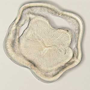
Figure D
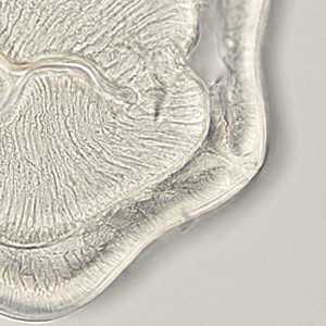
Figure E
Case Answer
This was a case of anisakiasis caused by Anisakis sp. The main diagnostic features shown in the images were:
- presence of three lips (Figure B).
- presence of Y-shaped lateral chords (black arrow, Figure E) and intestinal cells with a single basal nucleus (blue arrow, Figure E) and a brush border.
Figure B
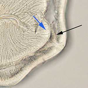
Figure E
More on: Anisakiasis
Images presented in the monthly case studies are from specimens submitted for diagnosis or archiving. On rare occasions, clinical histories given may be partly fictitious.
DPDx is an education resource designed for health professionals and laboratory scientists. For an overview including prevention and control visit www.cdc.gov/parasites/.
- Page last reviewed: August 24, 2016
- Page last updated: August 24, 2016
- Content source:
- Global Health – Division of Parasitic Diseases and Malaria
- Notice: Linking to a non-federal site does not constitute an endorsement by HHS, CDC or any of its employees of the sponsors or the information and products presented on the site.
- Maintained By: