


Case #400 - July 2015
 ShareCompartir
A ten-year-old boy presented at a local hospital with symptoms mimicking an appendicitis. An appendectomy was performed and biopsy specimens were sent to Pathology for routine histologic work-up. Objects suggestive of a parasite were observed by the attending pathologist. Figures A-E show what was observed on a slide stained with hematoxylin-and-eosin (H&E). What is your diagnosis? Based on what criteria?
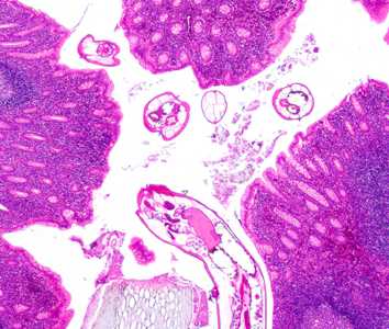
Figure A
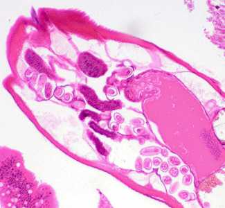
Figure B
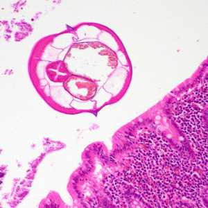
Figure C
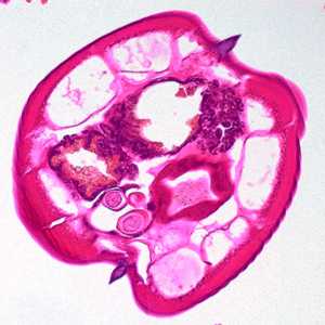
Figure D
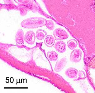
Figure E
Case Answer
This was a case of enterobiasis caused by the pinworm, Enterobius vermicularis. Diagnostic morphologic features included:
- platymyarian musculature (MU, Figure D) with relatively few cells per quadrant.
- prominent lateral alae (arrows, Figure D).
- elongate eggs (Figure E) with a thick shell that are within the size range for E. vermicularis (normal range 50-60 μm in length).
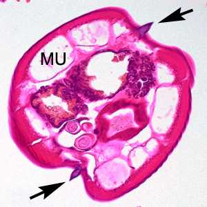
Figure D
Figure E
More on: Enterobiasis
This case and images were kindly provided by the University of Miami Health System/Jackson Memorial Hospital, Miami, FL.
Images presented in the monthly case studies are from specimens submitted for diagnosis or archiving. On rare occasions, clinical histories given may be partly fictitious.
DPDx is an education resource designed for health professionals and laboratory scientists. For an overview including prevention and control visit www.cdc.gov/parasites/.
- Page last reviewed: August 24, 2016
- Page last updated: August 24, 2016
- Content source:
- Global Health – Division of Parasitic Diseases and Malaria
- Notice: Linking to a non-federal site does not constitute an endorsement by HHS, CDC or any of its employees of the sponsors or the information and products presented on the site.
- Maintained By: