


Case #399 - July 2015
 ShareCompartir
A skin biopsy specimen was collected from the clavicle region of a 45-year-old male who presented with what appeared to be a pigmented lesion. The patient resides in Kentucky and has no known international travel. The biopsy specimen was sent to Pathology for routine histologic work-up. Objects suggestive of an organism were examined on slides stained with hematoxylin-and-eosin (H&E). The attending pathologist captured images and sent them to CDC-DPDx for diagnostic assistance. Figures A-E represent five of the images received. What is your diagnosis? Based on what criteria?
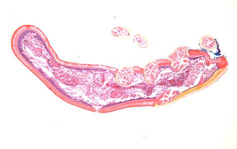
Figure A
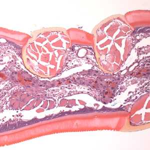
Figure B
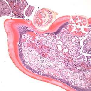
Figure C
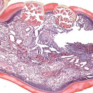
Figure D
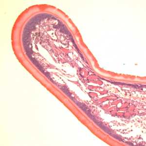
Figure E
Case Answer
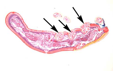
Figure A
More on: Ticks
This case and images were kindly provided by the Lexington Clinic, Lexington, KY.
Images presented in the monthly case studies are from specimens submitted for diagnosis or archiving. On rare occasions, clinical histories given may be partly fictitious.
DPDx is an education resource designed for health professionals and laboratory scientists. For an overview including prevention and control visit www.cdc.gov/parasites/.
- Page last reviewed: August 24, 2016
- Page last updated: August 24, 2016
- Content source:
- Global Health – Division of Parasitic Diseases and Malaria
- Notice: Linking to a non-federal site does not constitute an endorsement by HHS, CDC or any of its employees of the sponsors or the information and products presented on the site.
- Maintained By: