


Case #387 - January 2015
 ShareCompartir
A 71-year-old female presented to her health care provider with a nodule on her back. The nodule was removed and sent to Pathology for histological work-up. Tissue was sectioned, stained with hematoxylin-and-eosin (H & E) and examined by the attending pathologist and in turn forwarded to the CDC-DPDx for diagnostic assistance. A brief pathology report indicated a possible helminth infection. Figure A was taken at 100x, Figures B and C at 200x, and Figure D at 400x magnification. What is your diagnosis? Based on what criteria?
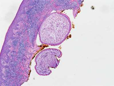
Figure A
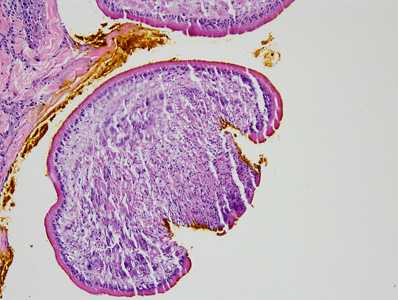
Figure B
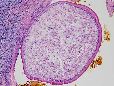
Figure C
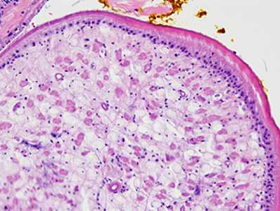
Figure D
Case Answer
This was a case of sparganosis caused by the third stage plerocercoid larva (sparganum) of a cestode in the genus Spirometra. Diagnostic features include:
- a thick tegument.
- the presence of calcareous corpuscles (best seen at 200x and 400x magnification).
- an absence of hooklets or defined protoscoleces helps to rule out echinococcosis, cysticercosis, and coenurosis.
Dogs, cats and other predatory mammals normally serve as the definitive host. Humans become accidental paratenic hosts by ingestion of water contaminated with infected copepods or consumption of raw or undercooked infected amphibians, reptiles, or fish.
More on: sparganosis
Images presented in the monthly case studies are from specimens submitted for diagnosis or archiving. On rare occasions, clinical histories given may be partly fictitious.
DPDx is an education resource designed for health professionals and laboratory scientists. For an overview including prevention and control visit www.cdc.gov/parasites/.
- Page last reviewed: August 24, 2016
- Page last updated: August 24, 2016
- Content source:
- Global Health – Division of Parasitic Diseases and Malaria
- Notice: Linking to a non-federal site does not constitute an endorsement by HHS, CDC or any of its employees of the sponsors or the information and products presented on the site.
- Maintained By: