


Case #379 - September 2014
 ShareCompartir
A 46-year-old woman returned from a trip to Nigeria with multiple boils on her lower back and extremities. Under the care of her primary physician, several fly larvae, one from each boil, were extracted and sent to the state health department for identification. The specimens were in turn forwarded to the CDC-DPDx for further diagnostic assistance. Figure A shows two of the larvae; Figure B shows a close-up of the posterior spiracles of one of the specimens. What is your identification? Based on what criteria?
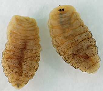
Figure A
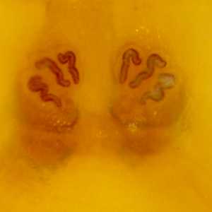
Figure B
Case Answer
This was a case of furuncular (cutaneous) myiasis caused by the tumbu fly, Cordylobia anthropophaga. The main diagnostic feature was the characteristic posterior spiracles (Figure B), which are represented by three sinuous slits each and no defined peritreme. The travel history was also helpful, as this species is endemic to sub-Saharan Africa. In most cases of myiasis, removal of the larvae is curative and species-level identification is not required for patient management.
More on: myiasis
Images presented in the monthly case studies are from specimens submitted for diagnosis or archiving. On rare occasions, clinical histories given may be partly fictitious.
DPDx is an education resource designed for health professionals and laboratory scientists. For an overview including prevention and control visit www.cdc.gov/parasites/.
- Page last reviewed: August 24, 2016
- Page last updated: August 24, 2016
- Content source:
- Global Health – Division of Parasitic Diseases and Malaria
- Notice: Linking to a non-federal site does not constitute an endorsement by HHS, CDC or any of its employees of the sponsors or the information and products presented on the site.
- Maintained By: