


Case #374 - July 2014
 ShareCompartir
A 34-year-old missionary worker sought medical attention for abdominal pain, nausea, and watery diarrhea after returning from visiting friends in Central America. A stool specimen was collected for laboratory testing and a modified Kinyoun’s acid-fast stained smear was prepared. An aliquot of the stool, along with the acid-fast stained smear, was sent to the CDC-DPDx laboratory for confirmatory testing. Figures A and B show what was observed in moderate numbers on the acid-fast stained smear at 500x magnification with oil. Figures C-F were taken at 400x magnification from a wet mount of the stool specimen. Figures C and E were taken using bright-field microscopy; Figures D and F represent the same fields, respectively, viewed using ultraviolet (UV) microscopy. The objects of interest measured 8-9 micrometers in diameter on average. What is your diagnosis? Based on what criteria?
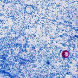
Figure A
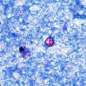
Figure B
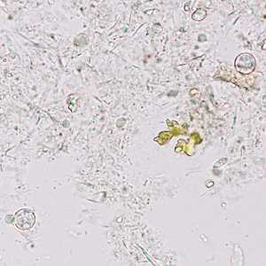
Figure C
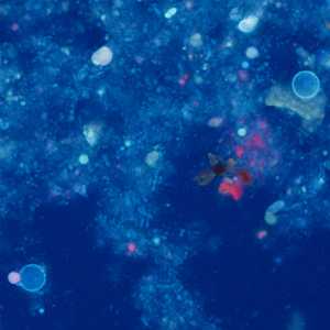
Figure D
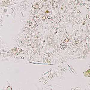
Figure E
Case Answer
This was a case of cyclosporiasis caused by Cyclospora cayetanensis. Diagnostic features included:
- round, refractile non-sporulated oocysts within the size range for Cyclospora (Figures C and E).
- oocysts whose wall autofluoresce when viewed with UV microscopy (Figures D and F).
- variable acid-fast staining oocysts (Figure A).
More on: cyclosporiasis
Images presented in the monthly case studies are from specimens submitted for diagnosis or archiving. On rare occasions, clinical histories given may be partly fictitious.
DPDx is an education resource designed for health professionals and laboratory scientists. For an overview including prevention and control visit www.cdc.gov/parasites/.
- Page last reviewed: August 24, 2016
- Page last updated: August 24, 2016
- Content source:
- Global Health – Division of Parasitic Diseases and Malaria
- Notice: Linking to a non-federal site does not constitute an endorsement by HHS, CDC or any of its employees of the sponsors or the information and products presented on the site.
- Maintained By: