


Case #373 - June 2014
 ShareCompartir
A worm-like object was observed during a routine colonoscopy screening of an adult living in Hawaii. The specimen was collected, sectioned, and stained with hematoxylin-and-eosin (H&E). Figures A-C show what was observed by the attending pathologist. Figure A was taken at 20x magnification; Figures B and C at 100x. What is your diagnosis? Based on what criteria?
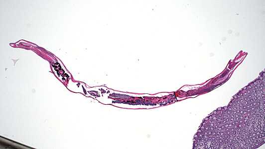
Figure A
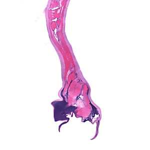
Figure B
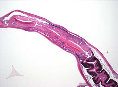
Figure C
Case Answer
This was a case of hookworm infection. Diagnostic features shown in the images included:
- a strong muscled esophagus (Figures A and C).
- presence of a buccal capsule (Figure A) characteristic of adult hookworms.
- the presence of a bursa (Figure B), which indicated that the worm was a male.
An adequate presentation of the buccal capsule was not available to determine if the worm was Necator or Ancylostoma.
More on: hookworm
Images presented in the monthly case studies are from specimens submitted for diagnosis or archiving. On rare occasions, clinical histories given may be partly fictitious.
DPDx is an education resource designed for health professionals and laboratory scientists. For an overview including prevention and control visit www.cdc.gov/parasites/.
- Page last reviewed: August 24, 2016
- Page last updated: August 24, 2016
- Content source:
- Global Health – Division of Parasitic Diseases and Malaria
- Notice: Linking to a non-federal site does not constitute an endorsement by HHS, CDC or any of its employees of the sponsors or the information and products presented on the site.
- Maintained By: