


Case #362 - December 2013
 ShareCompartir
A 29-year-old female post-graduate student in Zoology went on an expedition to see the lowland gorillas in the Democratic Republic of Congo. She reported numerous insect bites while traveling but did take anti-malarial prophylaxis. Approximately one week after returning home she developed fever. About a month later, she started experiencing headaches, itchy skin, and swollen lymph nodes and sought medical attention. A blood specimen was collected; smears made and stained with Wright-Giemsa. The objects shown in Figures A-D were observed in moderate numbers. What is your diagnosis? Based on what criteria?
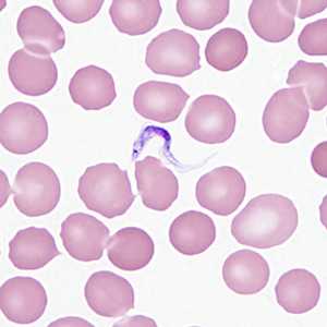
Figure A
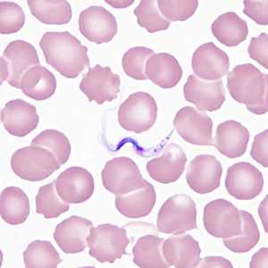
Figure B
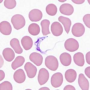
Figure C
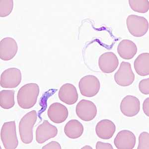
Figure D
Case Answer
This was a case of African trypanosomiasis caused by a hemoflagellate belonging to the Trypanosoma brucei complex. Diagnostic morphologic features shown included:
- trypomastigotes with a centrally located nucleus, an undulating membrane, and a flagellum running along the undulating membrane.
- a small kinetoplast located on the anterior end of the trypomastigote.
- dividing forms (Figures C and D).
This was most likely T. b. gambiense (although morphologically it is identical to T. b. rhodesiense), given the travel history to the DRC.
More on: African trypanosomiasi
Images presented in the monthly case studies are from specimens submitted for diagnosis or archiving. On rare occasions, clinical histories given may be partly fictitious.
DPDx is an education resource designed for health professionals and laboratory scientists. For an overview including prevention and control visit www.cdc.gov/parasites/.
- Page last reviewed: August 24, 2016
- Page last updated: August 24, 2016
- Content source:
- Global Health – Division of Parasitic Diseases and Malaria
- Notice: Linking to a non-federal site does not constitute an endorsement by HHS, CDC or any of its employees of the sponsors or the information and products presented on the site.
- Maintained By: