


Case #350 - June, 2013
 ShareCompartir
A 45-year-old man with travel history to Ghana presented to his health care provider with recurrent fever and chills. He admitted to not taking any kind of malaria prophylaxis during his travels. Blood smears were prepared, stained with Wright's stain, and examined. The laboratory initially reported Plasmodium falciparum but requested confirmation from a reference laboratory, so the slides and EDTA blood were sent to the CDC-DPDx for diagnostic assistance. Figures A-I show what was found on one of the stained thin smears. What is your diagnosis? Based on what criteria? What, if any, other tests would you recommend?
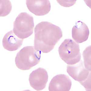
Figure A
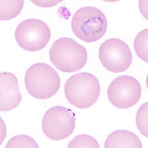
Figure B
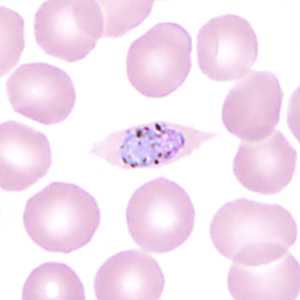
Figure C
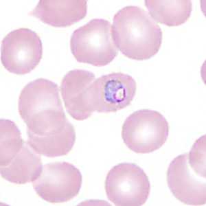
Figure D
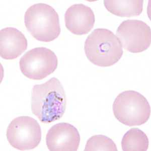
Figure E
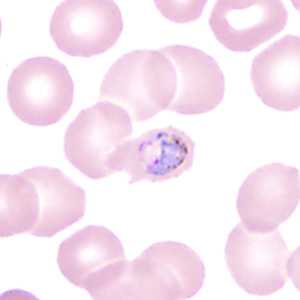
Figure F
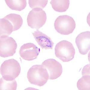
Figure G
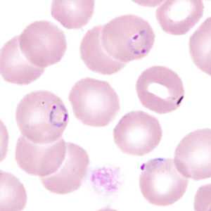
Figure H
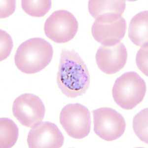
Figure I
Case Answer
This was a case of malaria caused by two species of Plasmodium, P. falciparum and P. ovale. Diagnostic features:
- thin, delicate rings in normal-sized RBCs, some with double chromatin dots (Figures A, B, E, G, and H).
- appliqué forms (Figures A, B, G, and H) (although this feature is not exclusive to P. falciparum, the morphology of these particular rings and the infected RBC is still more consistent with P. falciparum).
- enlarged and/or oval-shaped infected RBCs (Figures B, C, D, E, F, G, and I) consistent with P. ovale.
- infected RBCs showing fimbriation (Figures C, D, E, F, G, and I) consistent with P. ovale.
- trophozoites in enlarged infected RBCs (Figures B, D, F, and G) consistent with P. ovale.
- gametocytes in enlarged infected RBCs (Figures C, E, and I) with coarse pigment consistent with P. ovale.
Real-time PCR was performed on the EDTA blood and confirmed a mixed infection with the two species of Plasmodium. It should be noted that blood smears stained with Wright's or Wright-Giemsa often may not show Schüffner's stippling, and that other morphologic features may still be used to make a species-level identification. PCR should always be used when there is morphologic evidence of a mixed infection.
More on: Malaria
Images presented in the monthly case studies are from specimens submitted for diagnosis or archiving. On rare occasions, clinical histories given may be partly fictitious.
DPDx is an education resource designed for health professionals and laboratory scientists. For an overview including prevention and control visit www.cdc.gov/parasites/.
- Page last reviewed: August 24, 2016
- Page last updated: August 24, 2016
- Content source:
- Global Health – Division of Parasitic Diseases and Malaria
- Notice: Linking to a non-federal site does not constitute an endorsement by HHS, CDC or any of its employees of the sponsors or the information and products presented on the site.
- Maintained By: