


Case #344 - March, 2013
 ShareCompartir
A 26-year-old female presented at a local hospital with severe pain and bloody discharge from the ear. The symptoms started while on a returning flight from vacation in Central America. The patient explained that while on vacation she visited a local physician to have a fly removed from her ear canal. Tissue was removed and sent to Pathology for testing. Several viable fly larvae were observed in the tissue. The case was diagnosed as aural myiasis and the larvae were allowed to mature to aid in identification. Images of mature (third instar) larvae were captured and sent to the DPDx Team for diagnostic assistance. Figure A shows one of the larvae; Figure B shows a close-up of its posterior spiracles. What is your identification? Based on what criteria?
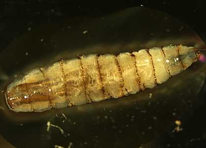
Figure A
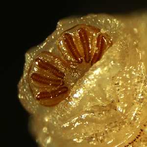
Figure B
Case Answer
This was a case of aural myiasis caused by the primary screwworm fly, Cochliomyia hominovorax. Diagnostic morphologic features included:
- an elongate, slightly tapering habitus with cuticular spines on each body segment (Figure A).
- posterior spiracles with straight slits, a weakly-sclerotized peritreme with lateral swellings, and an obsolete to absent ecdysial scar (button) (Figure B).
Travel was also supportive, as C. hominovorax is endemic to tropical America. While eggs are usually laid in wounds, C. hominovorax will attack healthy tissue and can be damaging to the host if not removed promptly.
More on: Myiasis
This case and images were kindly provided by the University of Washington Medical Center, Seattle, WA.
Images presented in the monthly case studies are from specimens submitted for diagnosis or archiving. On rare occasions, clinical histories given may be partly fictitious.
DPDx is an education resource designed for health professionals and laboratory scientists. For an overview including prevention and control visit www.cdc.gov/parasites/.
- Page last reviewed: August 24, 2016
- Page last updated: August 24, 2016
- Content source:
- Global Health – Division of Parasitic Diseases and Malaria
- Notice: Linking to a non-federal site does not constitute an endorsement by HHS, CDC or any of its employees of the sponsors or the information and products presented on the site.
- Maintained By: