


Case #338 - December, 2012
 ShareCompartir
A 42-year-old pig farmer presented to his local medical facility with intermittent diarrhea and abdominal cramping. A stool specimen was collected in 10% formalin for laboratory testing. Figures A and C were captured at 200x magnification and show what was found in high numbers. Figures B, D, and E show a higher magnification (400x) of the same objects. What is your diagnosis? Based on what criteria?
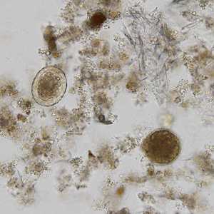
Figure A
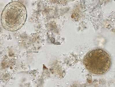
Figure B
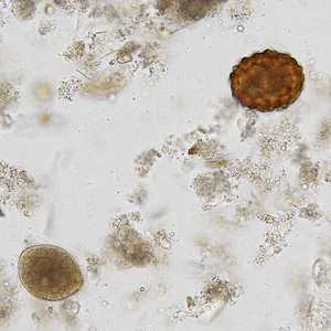
Figure C
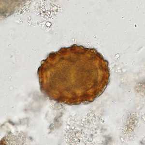
Figure D
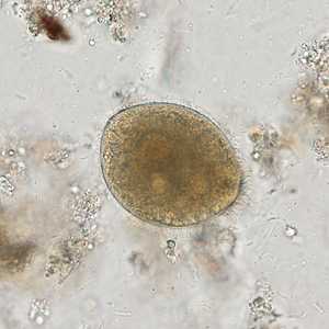
Figure E
Case Answer
This was a mixed infection with Ascaris lumbricoides and Balantidium coli. Morphologic features shown in the figures included:
- unembryonated, fertile eggs of A. lumbricoides demonstrating a thick wall (Figures A-D) and with a mammillated layer (Figures C and D).
- large smooth-walled cyst of Balantidium coli (Figures A and B).
- trophozoite of B. coli demonstrating cilia, a large macronucleus, and a cytostome (Figures C and E).
More on: Ascariasis; Balantidiasis
Images presented in the monthly case studies are from specimens submitted for diagnosis or archiving. On rare occasions, clinical histories given may be partly fictitious.
DPDx is an education resource designed for health professionals and laboratory scientists. For an overview including prevention and control visit www.cdc.gov/parasites/.
- Page last reviewed: August 24, 2016
- Page last updated: August 24, 2016
- Content source:
- Global Health – Division of Parasitic Diseases and Malaria
- Notice: Linking to a non-federal site does not constitute an endorsement by HHS, CDC or any of its employees of the sponsors or the information and products presented on the site.
- Maintained By: