


Case #324 - May, 2012
 ShareCompartir
A 58-year-old female who had emigrated from Africa was seen by her health care provider for complications with squamous cell carcinoma of the cervix. CT scans of the pelvis revealed a small mass in the bladder. Biopsy specimens of the bladder and subsequent bladder washings were sent to Pathology for histological work-up. Microscopic examination of the specimens by the attending pathologist revealed structures suggestive of a parasite. Digital images were sent to the DPDx Team for diagnostic assistance. Figures A-D show what was observed in the biopsy specimens, stained with hematoxylin and eosin (H&E). Figures E and F show what was observed in the bladder washings, stained with Papanicolaou stain. The object in Figure E measured 150 micrometers in length. What is your diagnosis? Based on what criteria?
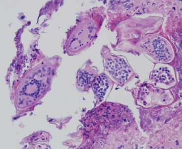
Figure A
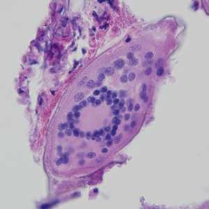
Figure B
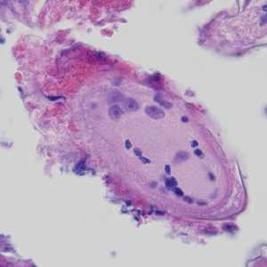
Figure C

Figure D
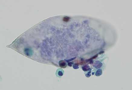
Figure E
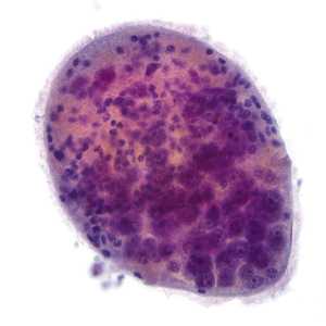
Figure F
Case Answer
This was a case of schistosomiasis caused by Schistosoma haematobium. Morphologic diagnostic features included:
- presence of eggs in bladder tissue (Figures A-D).
- elongated, non-operculated eggs bearing a terminal spine and within the size range for S. haematobium (Figure E).
- eggs containing defined miracidia (best seen in the biopsy sections in Figures B-D).
- free ciliated miracidium (Figure F).
More on: Schistosomiasis
Images presented in the monthly case studies are from specimens submitted for diagnosis or archiving. On rare occasions, clinical histories given may be partly fictitious.
DPDx is an education resource designed for health professionals and laboratory scientists. For an overview including prevention and control visit www.cdc.gov/parasites/.
- Page last reviewed: August 24, 2016
- Page last updated: August 24, 2016
- Content source:
- Global Health – Division of Parasitic Diseases and Malaria
- Notice: Linking to a non-federal site does not constitute an endorsement by HHS, CDC or any of its employees of the sponsors or the information and products presented on the site.
- Maintained By: