


Case #311 - November, 2011
 ShareCompartir
A 55-year-old homebound Type II diabetic, originally from Haiti, was hospitalized for severe abdominal pain and hematemesis. Blood specimens were collected and sent to Hematology for routine work-up. Digital images were captured on a Wright-stained thin smear from a blood specimen collected at 2:25AM. The images (Figure A-C) were sent via email to the DPDx Team for diagnostic assistance. Measurements were not included in the email. What is your diagnosis? Based on what criteria?
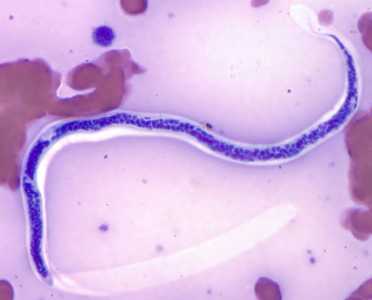
Figure A
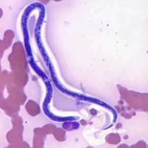
Figure B
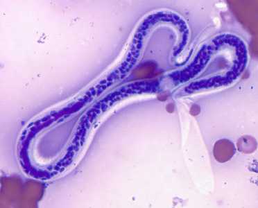
Figure C
Case Answer
This was a case of Bancroft's filariasis caused by Wuchereria bancrofti. Diagnostic criteria included:
- sheathed microfilariae with an unstained sheath.
- microfilariae with a relatively short head space and an anucleate tail.
- nocturnal periodicity.
- history of residence in known area endemic for W. bancrofti.
More on: Lympthatic Filariasis
Images presented in the monthly case studies are from specimens submitted for diagnosis or archiving. On rare occasions, clinical histories given may be partly fictitious.
DPDx is an education resource designed for health professionals and laboratory scientists. For an overview including prevention and control visit www.cdc.gov/parasites/.
- Page last reviewed: August 24, 2016
- Page last updated: August 24, 2016
- Content source:
- Global Health – Division of Parasitic Diseases and Malaria
- Notice: Linking to a non-federal site does not constitute an endorsement by HHS, CDC or any of its employees of the sponsors or the information and products presented on the site.
- Maintained By: