


Case #283 - September, 2010
 ShareCompartir
A 49-year-old man presented to his primary care physician with a lump in his throat which had developed over the past three months. He was ultimately referred to an oral surgeon with multiple oral ulcers and submucosal nodules. The patient was originally from Mexico, but has lived in Iowa for nearly 20 years, with recent visits back to Mexico in the past two years. The patient currently works as a landscaper, but has also worked in a foundry and on a pig farm. Biopsy specimens were taken from the submucosal nodules and sent to a pathology laboratory for routine histologic sectioning. Figures A-F show what was observed in sections of the nodule, stained with hematoxylin and eosin (H&E). Figure A was taken at 100x magnification. Figures B and C were taken at 400x magnification. Figures D-F were taken at 1000x magnification. What is your diagnosis? Based on what criteria?
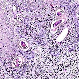
Figure A
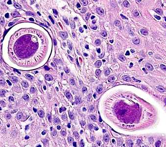
Figure B
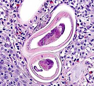
Figure C
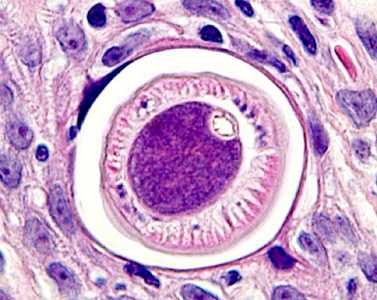
Figure D
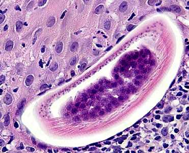
Figure E
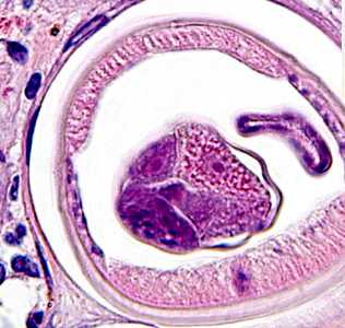
Figure F
Case Answer
This was a case of anatrichosomiasis caused by a trichuroid nematode in the genus, Anatrichosoma (possibly A. buccalis given travel history and clinical manifestation). Diagnostic morphologic features included:
- tall, polymyarian musculature (MU, Figure D).
- the presence of bacillary bands, a characteristic of trichuroid nematodes (BB, Figure D).
- a small esophagus (ES, Figure D), embedded in a stichocyte (ST, Figure D).
- infertile eggs within the single reproductive tube (EG, Figure F).
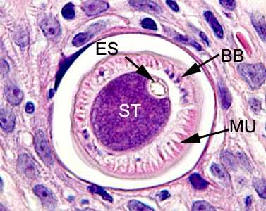
Figure D
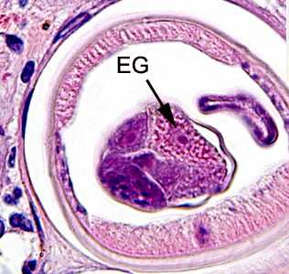
Figure F
Anatrichosoma buccalis in nature is a parasite found in the oral cavity of opossums in North America. Very little is known about the biology, life cycle and host range of species of Anatrichosoma. Eggs are shed embryonated in the environment but it is unclear if there is an intermediate host involved in the life cycle. There have been only a few human cases documented, most of them in subcutaneous skin nodules or breast nodules.
Images presented in the monthly case studies are from specimens submitted for diagnosis or archiving. On rare occasions, clinical histories given may be partly fictitious.
DPDx is an education resource designed for health professionals and laboratory scientists. For an overview including prevention and control visit www.cdc.gov/parasites/.
- Page last reviewed: August 24, 2016
- Page last updated: August 24, 2016
- Content source:
- Global Health – Division of Parasitic Diseases and Malaria
- Notice: Linking to a non-federal site does not constitute an endorsement by HHS, CDC or any of its employees of the sponsors or the information and products presented on the site.
- Maintained By: