


Case #282 - August, 2010
 ShareCompartir
A 49-year-old man with AIDS was admitted to the hospital for complaints of a persisting fever and dry cough. A chest radiograph showed bilateral infiltrate. A sputum specimen was collected and stained with Giemsa, but no parasites were observed. A lung biopsy was obtained, sectioned, and stained with both methenamine silver and hematoxylin and eosin (H&E) stains. Figures A and B show what was observed at 500x magnification on one of the stained sections. What is your diagnosis? Based on what criteria?
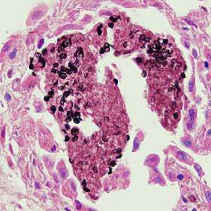
Figure A
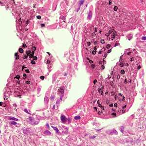
Figure B
Case Answer
This was a case of pneumonia caused by Pneumocystis jirovecii (previously classified as Pneumocystis carinii). Because this case showed images taken from a lung biopsy stained with a combination of hematoxylin and eosin (H&E) and methenamine silver, only the cyst walls stained black and intracystic bodies are not visible.
More on: Pneumocystis jirovecii
Images presented in the monthly case studies are from specimens submitted for diagnosis or archiving. On rare occasions, clinical histories given may be partly fictitious.
DPDx is an education resource designed for health professionals and laboratory scientists. For an overview including prevention and control visit www.cdc.gov/parasites/.
- Page last reviewed: August 24, 2016
- Page last updated: August 24, 2016
- Content source:
- Global Health – Division of Parasitic Diseases and Malaria
- Notice: Linking to a non-federal site does not constitute an endorsement by HHS, CDC or any of its employees of the sponsors or the information and products presented on the site.
- Maintained By: