


Case #281 - August, 2010
 ShareCompartir
A twenty-year-old male from India presented with recurrent abdominal pain. He underwent an appendectomy at a local medical center. Sections of the appendix were obtained, sectioned, and stained with hematoxylin and eosin (H&E). Figures A–D show what was observed microscopically. What is your diagnosis? Based on what criteria?
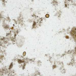
Figure A
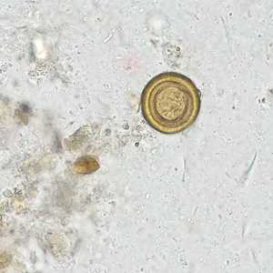
Figure B
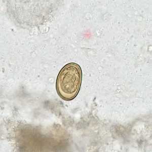
Figure C
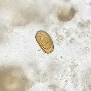
Figure D
Case Answer
This was a case of enterobiasis caused by Enterobius vermicularis (pinworm). Morphologic diagnostic features shown included:
- the presence of platymyarian muscle cells (Figure A).
- prominent lateral alae (Figures A, B, and C).
- embryonated eggs in utero that were flattened on one side (Figures C and D).
More on: Enterobiasis
This case and images were kindly provided by Dr. CSBR Prasad, Dept. of Pathology, Sri Devaraj Urs Medical College, Karnataka, India.
Images presented in the monthly case studies are from specimens submitted for diagnosis or archiving. On rare occasions, clinical histories given may be partly fictitious.
DPDx is an education resource designed for health professionals and laboratory scientists. For an overview including prevention and control visit www.cdc.gov/parasites/.
- Page last reviewed: August 24, 2016
- Page last updated: August 24, 2016
- Content source:
- Global Health – Division of Parasitic Diseases and Malaria
- Notice: Linking to a non-federal site does not constitute an endorsement by HHS, CDC or any of its employees of the sponsors or the information and products presented on the site.
- Maintained By: