


Case #274 - April, 2010
 ShareCompartir
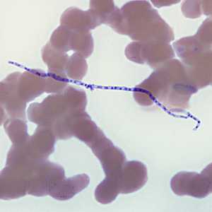
Figure A
Case Answer
The objects in this case were artifacts (fungal elements, possibly Helicosporium sp. or related) and a diagnosis was made of No Parasites Found (NPF). Fungal hyphae and conidia are not uncommon airborne contaminants in the laboratory. If such objects were to alight on a drying blood smear, they could get stained, observed and possibly misinterpreted as parasites. In this case, the objects most-closely resembled a microfilaria. Careful examination of morphologic features, as well as accurate measurements, should be performed to ensure a proper diagnosis.
More on: Artifacts
Images presented in the monthly case studies are from specimens submitted for diagnosis or archiving. On rare occasions, clinical histories given may be partly fictitious.
DPDx is an education resource designed for health professionals and laboratory scientists. For an overview including prevention and control visit www.cdc.gov/parasites/.
- Page last reviewed: August 24, 2016
- Page last updated: August 24, 2016
- Content source:
- Global Health – Division of Parasitic Diseases and Malaria
- Notice: Linking to a non-federal site does not constitute an endorsement by HHS, CDC or any of its employees of the sponsors or the information and products presented on the site.
- Maintained By: