


Case #272 - March, 2010
 ShareCompartir
A 35-year-old male from the United Arab Emirates was hospitalized with complaints of upper and lower abdominal pain. Sections of the greater omentum and appendix were biopsied and sent to a pathology laboratory for sectioning and staining. Figures A-D show what was observed by the attending pathologist in sections of the greater omentum, stained with hematoxylin and eosin (H&E). What is your diagnosis? Based on what criteria? What other testing, if any, would you recommend?
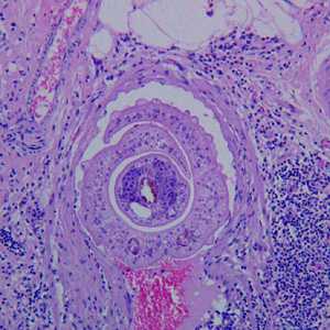
Figure A
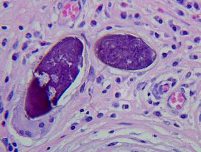
Figure B
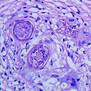
Figure C
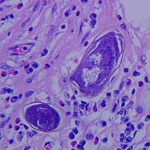
Figure D
Case Answer
This was a case of schistosomiasis, caused by Schistosoma mansoni. Diagnostic morphologic features included:
- cross-sections of adult worms (Figure A), with the female residing in the gynecophoral canal of the male.
- cross-sections of eggs showing miracidia (Figure C).
- transverse-sections of eggs (Figures B and D), one of which (Figure D) shows the lateral spine characteristic of S. mansoni.
Although a species-level identification could be made by the images in this case, serology is also recommended when schistosomiasis is suspected.
More on: Schistosomiasis
This case and images were kindly provided by Dr. Munaf Desai, Al Qassimi Hospital, Shatjah, UAE.
Images presented in the monthly case studies are from specimens submitted for diagnosis or archiving. On rare occasions, clinical histories given may be partly fictitious.
DPDx is an education resource designed for health professionals and laboratory scientists. For an overview including prevention and control visit www.cdc.gov/parasites/.
- Page last reviewed: August 24, 2016
- Page last updated: August 24, 2016
- Content source:
- Global Health – Division of Parasitic Diseases and Malaria
- Notice: Linking to a non-federal site does not constitute an endorsement by HHS, CDC or any of its employees of the sponsors or the information and products presented on the site.
- Maintained By: