


Case #271 - March, 2010
 ShareCompartir
The DPDx Team received Giemsa-stained blood smears from a state health laboratory for malaria confirmation/identification. The patient travels internationally regularly for business, and within the month before becoming ill, had traveled to India, Malaysia and China. Figures A-F show what was observed on one of the thin smears sent to the CDC. What is your diagnosis? Based on what criteria?
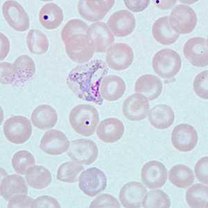
Figure A
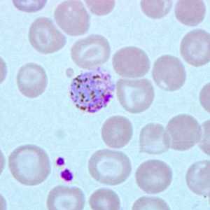
Figure B
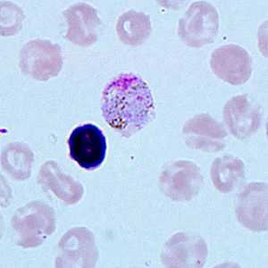
Figure C
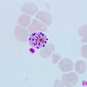
Figure D
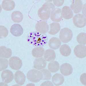
Figure E
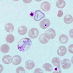
Figure F
Case Answer
This was a case of malaria caused by Plasmodium vivax. Diagnostic morphologic features included:
- an enlargement of infected red blood cells.
- the presence of Schϋffner's dots (best seen in Figures A and F).
- amoeboid trophozoites (Figures A and F).
- gametocytes with fine pigment (Figures B and C).
- schizonts with more than 13 merozoites (Figures D and E).
More on: Malaria
Images presented in the monthly case studies are from specimens submitted for diagnosis or archiving. On rare occasions, clinical histories given may be partly fictitious.
DPDx is an education resource designed for health professionals and laboratory scientists. For an overview including prevention and control visit www.cdc.gov/parasites/.
- Page last reviewed: August 24, 2016
- Page last updated: August 24, 2016
- Content source:
- Global Health – Division of Parasitic Diseases and Malaria
- Notice: Linking to a non-federal site does not constitute an endorsement by HHS, CDC or any of its employees of the sponsors or the information and products presented on the site.
- Maintained By: