


Case #270 - March, 2010
 ShareCompartir
A six-year-old boy was taken to a local clinic for abdominal discomfort. A stool specimen was collected in formalin for routine ova-and-parasite (O&P) examination. Figures A-B show what was observed on an unstained wet mount preparation after the specimen was concentrated. All images were taken at 400x magnification. The objects of interest measured approximately 40-43 micrometers in diameter. What is your diagnosis? Based on what criteria?
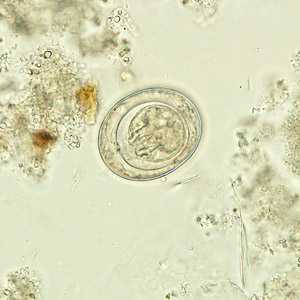
Figure A
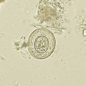
Figure B
Case Answer
This was a case of hymenolepiasis caused by Hymenolepis nana (the dwarf tapeworm). Diagnostic morphologic features shown in the figures included:
- eggs with a thin, hyaline shell and an oncosphere in which hooks can be seen. Six hooks should be present, but they are not always visible in the same focal plane.
- eggs within a size range consistent with those of H. nana (30 to 50 micrometers in diameter). Size is the primary feature used to distinguish eggs of H. nana from those of H. diminuta (70-85 micrometers by 60-80 micrometers).
- the presence of polar filaments extending from opposite ends of the oncosphere (lacking in H. diminuta).
More on: Hymenolepiasis
Images presented in the monthly case studies are from specimens submitted for diagnosis or archiving. On rare occasions, clinical histories given may be partly fictitious.
DPDx is an education resource designed for health professionals and laboratory scientists. For an overview including prevention and control visit www.cdc.gov/parasites/.
- Page last reviewed: August 24, 2016
- Page last updated: August 24, 2016
- Content source:
- Global Health – Division of Parasitic Diseases and Malaria
- Notice: Linking to a non-federal site does not constitute an endorsement by HHS, CDC or any of its employees of the sponsors or the information and products presented on the site.
- Maintained By: