


Case #262 - October, 2009
 ShareCompartir
A 70-year-old man from the mid-western United States had complaints of fever and general malaise lasting for about one week. He was admitted to a hospital with evidence of hemolytic process (dark urine and low hemoglobin). He had undergone a splenectomy when he was 40 years old. Blood smears were ordered, prepared, and stained with Giemsa. Figures A and B show what was observed on the stained smears at 1000x magnification. What is your diagnosis? Based on what criteria? What other testing, if any, would you recommend?
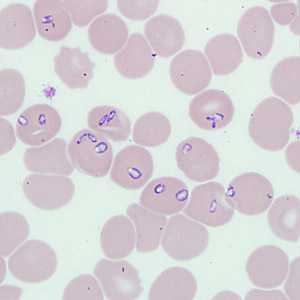
Figure A
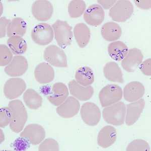
Figure B
Case Answer
This was a case of babesiosis caused by Babesia sp. Diagnostic morphologic features shown in Figures A and B were:
- pleomorphic and vacuolated intraerythrocytic ring-like parasites in normal-sized red blood cells.
- tetrad formations.
- an absence of the pigment which can be found in Plasmodium species.
In general, identification of Babesia to the species or strain level is not possible by morphology, and requires molecular analysis of a blood specimen. PCR was used to confirm that the parasite identified in this specimen was the species of Babesia currently designated MO-1.
More on: Babesiosis
Images presented in the monthly case studies are from specimens submitted for diagnosis or archiving. On rare occasions, clinical histories given may be partly fictitious.
DPDx is an education resource designed for health professionals and laboratory scientists. For an overview including prevention and control visit www.cdc.gov/parasites/.
- Page last reviewed: August 24, 2016
- Page last updated: August 24, 2016
- Content source:
- Global Health – Division of Parasitic Diseases and Malaria
- Notice: Linking to a non-federal site does not constitute an endorsement by HHS, CDC or any of its employees of the sponsors or the information and products presented on the site.
- Maintained By: