


Case #255 - July, 2009
 ShareCompartir
A 36-year-old man was admitted to the hospital with fever, chills, headache, and myalgia. The patient recently returned to the United States following a month-long trip to Rwanda to visit family. Blood was collected in EDTA and sent to the Hematology Department for work-up. Figures A-F show what was observed on a thin smear made from the blood, stained with Giemsa. All images were captured at 1000x magnification. What is your diagnosis? Based on what criteria?
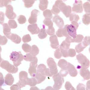
Figure A
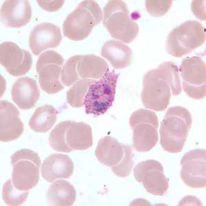
Figure B
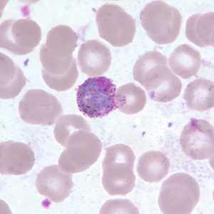
Figure C
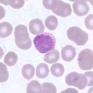
Figure D
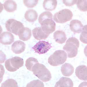
Figure E
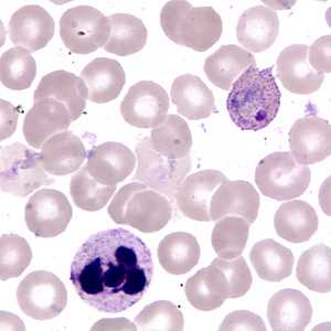
Figure F
Case Answer
This was a case of malaria caused by Plasmodium ovale. Diagnostic features included:
- infected RBCs showing slight enlargement.
- infected RBCs with trophozoites exhibiting elongation, fimbriation and Schüffner's stippling (Figures A, B, E, and F).
- infected RBCs with macrogametocytes that fill the host RBC and exhibit coarse pigment, compact chromatin, and Schüffner's stippling (Figures C and D).
More on: Malaria
Images presented in the monthly case studies are from specimens submitted for diagnosis or archiving. On rare occasions, clinical histories given may be partly fictitious.
DPDx is an education resource designed for health professionals and laboratory scientists. For an overview including prevention and control visit www.cdc.gov/parasites/.
- Page last reviewed: August 24, 2016
- Page last updated: August 24, 2016
- Content source:
- Global Health – Division of Parasitic Diseases and Malaria
- Notice: Linking to a non-federal site does not constitute an endorsement by HHS, CDC or any of its employees of the sponsors or the information and products presented on the site.
- Maintained By: