


Case #253 - June, 2009
 ShareCompartir
A 30-month-old child born in China was recently adopted by a family in the Netherlands. Three consecutive stool specimens were obtained for ova and parasite (O&P) testing, as part of the standard procedure during child adoption. There were no complaints. A formalin-ethyl acetate concentration and a water sedimentation method were performed. The following figures show what was observed on a wet mount prepared from the concentrate. Figure A-C were taken at 400x magnification; Figure D at 630x magnification. The objects were seen in moderate numbers in each concentrate and in all three stool specimens. The objects in Figure A measured approximately 24 to 37 micrometers. The objects in Figures B-D measured 73 to 85 micrometers in length by 44 micrometers in width. What is your diagnosis? Based on what criteria?
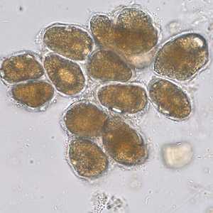
Figure A
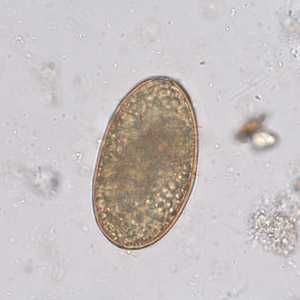
Figure B
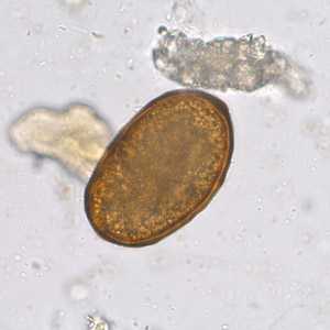
Figure C
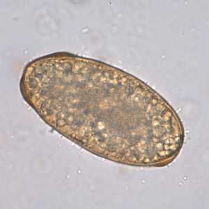
Figure D
Case Answer
This was a case of ascariasis caused by Ascaris lumbricoides. Figure A showed a conglomerate of plant cells. Figures B-D showed infertile, decorticated Ascaris eggs (usual size range 85-95 micrometers long by 40-50 micrometers wide).
More on: Ascariasis
Images presented in the monthly case studies are from specimens submitted for diagnosis or archiving. On rare occasions, clinical histories given may be partly fictitious.
DPDx is an education resource designed for health professionals and laboratory scientists. For an overview including prevention and control visit www.cdc.gov/parasites/.
- Page last reviewed: August 24, 2016
- Page last updated: August 24, 2016
- Content source:
- Global Health – Division of Parasitic Diseases and Malaria
- Notice: Linking to a non-federal site does not constitute an endorsement by HHS, CDC or any of its employees of the sponsors or the information and products presented on the site.
- Maintained By: