


Case #252 - May, 2009
 ShareCompartir
A 29-year-old man from Haiti had blood drawn for routine work-up, following hospitalization for complications with a congenital disorder. The blood was collected in EDTA tubes and sent to Hematology and Microbiology for evaluation. The following images show what was observed on a Giemsa-stained thick smear of the blood. The object measured 270 micrometers in length. The image in Figure A was captured at 500x magnification; the images in Figures B and C, which show close-ups of the two ends of the same object, were taken at 1000x magnification. What is your diagnosis? Based on what criteria?
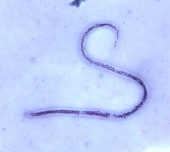
Figure A
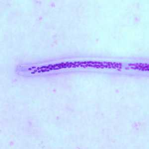
Figure B
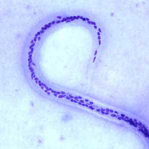
Figure C
Case Answer
This was a case of filariasis caused by Wuchereria bancrofti. Diagnostic features included:
- a microfilaria within the size range (240-300 micrometers) for W. bancrofti.
- a loosely-packed nuclear column.
- a tail that tapers to a point, with nuclei terminating before the end of the tail (Figure C).
Although the specimen presented in this case was unsheathed (microfilariae of W. bancrofti usually possess a sheath), the size, nuclear structure, and travel history should make for an accurate diagnosis.
More on: Lymphatic Filariasis
Images presented in the monthly case studies are from specimens submitted for diagnosis or archiving. On rare occasions, clinical histories given may be partly fictitious.
DPDx is an education resource designed for health professionals and laboratory scientists. For an overview including prevention and control visit www.cdc.gov/parasites/.
- Page last reviewed: August 24, 2016
- Page last updated: August 24, 2016
- Content source:
- Global Health – Division of Parasitic Diseases and Malaria
- Notice: Linking to a non-federal site does not constitute an endorsement by HHS, CDC or any of its employees of the sponsors or the information and products presented on the site.
- Maintained By: