


Case #251 - May, 2009
 ShareCompartir
A 45-year-old pig farmer from rural Georgia presented to his health care provider with abdominal pain, cramps and diarrhea. Stool specimens were collected in polyvinyl alcohol (PVA) and 10% formalin for ova and parasite (O&P) examination. The objects in Figures A and B, which measured on average 90 micrometers in length, were observed in low numbers in formalin-concentrated wet mounts of the stool. The objects in Figures C and D, which measured on average 12 micrometers in diameter, were seen in moderate numbers on trichrome-stained slides prepared from the PVA-preserved stool. What is your diagnosis? Based on what criteria?
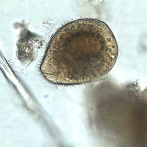
Figure A
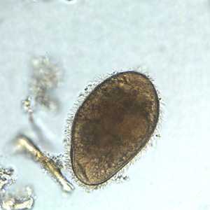
Figure B
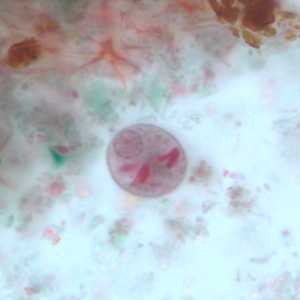
Figure C
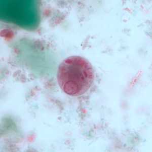
Figure D
Case Answer
This was a case of balantidiasis, caused by Balantidium coli (Figures A and B); also observed were cysts of Entamoeba polecki (Figures C and D). Diagnostic features included:
- ciliated trophozoites of B. coli (Figures A and B) containing a cytosome and macronucleus, and within the size range (40-200 micrometers) of the species.
- uninucleate cysts of E. polecki (Figures D and E) containing a nucleus with a pleomorphic karyosome and evenly-distributed peripheral chromatin, and many irregularly-shaped chromatoid bodies, as well as being within the size range (11-15 micrometers) of the species.
More on: Balantidiasis: Entamoeba polecki
Images presented in the monthly case studies are from specimens submitted for diagnosis or archiving. On rare occasions, clinical histories given may be partly fictitious.
DPDx is an education resource designed for health professionals and laboratory scientists. For an overview including prevention and control visit www.cdc.gov/parasites/.
- Page last reviewed: August 24, 2016
- Page last updated: August 24, 2016
- Content source:
- Global Health – Division of Parasitic Diseases and Malaria
- Notice: Linking to a non-federal site does not constitute an endorsement by HHS, CDC or any of its employees of the sponsors or the information and products presented on the site.
- Maintained By: