


Case #249 - April, 2009
 ShareCompartir
A small worm approximately 2.5 cm in length was recovered from the stomach of a 38-year-old male. The anterior end had been damaged by the extraction but it was sent to the CDC to see if it could still be identified. It was placed in lacto-phenol for clearing, after which it was still difficult to make a definitive identification. A small cross-section was made and examined using a compound microscope. The specimen in Figure A was captured at 40x magnification using brightfield illumination; the specimen in Figure B at 200x using differential interference contrast (DIC) microscopy. What is your diagnosis? Based on what criteria?
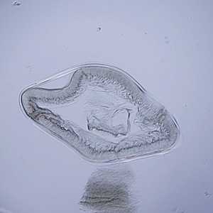
Figure A
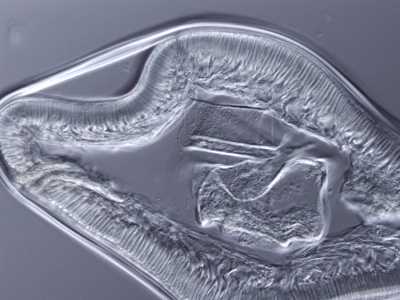
Figure B
Case Answer
This was a case of anisakiasis caused by Anisakis sp. The main diagnostic features shown in the images were:
- large Y-shaped lateral chords (black arrow, Figure B).
- tall, prominent muscle cells (red arrow, Figure B).
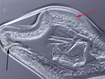
Figure B
More on: Anisakiasis
Images presented in the monthly case studies are from specimens submitted for diagnosis or archiving. On rare occasions, clinical histories given may be partly fictitious.
DPDx is an education resource designed for health professionals and laboratory scientists. For an overview including prevention and control visit www.cdc.gov/parasites/.
- Page last reviewed: August 24, 2016
- Page last updated: August 24, 2016
- Content source:
- Global Health – Division of Parasitic Diseases and Malaria
- Notice: Linking to a non-federal site does not constitute an endorsement by HHS, CDC or any of its employees of the sponsors or the information and products presented on the site.
- Maintained By: