


Case #243 - January, 2009
 ShareCompartir
A ten-year-old child returned from summer camp with abdominal discomfort, nausea and diarrhea. He was taken to his pediatrician and a stool specimen was collected for routine work-up, including ova and parasite (O&P) examination. Figures A-D show what was observed on a trichrome-stained slide prepared from a PVA-preserved aliquot of the stool. The objects in Figures A and B measured approximately 11 micrometers; the objects in Figures C and D measured approximately seven micrometers. What is your diagnosis? Based on what criteria?
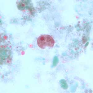
Figure A
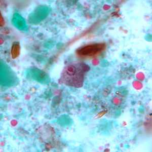
Figure B
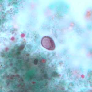
Figure C
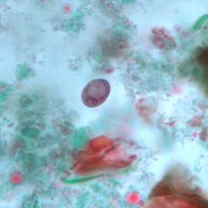
Figure D
Case Answer
These images showed the trophozoites and cysts of the nonpathogenic ameba, Iodamoeba buetschlii. Diagnostic features included:
- trophozoites (Figures A and B) within the size range (8-20 micrometers) for the species, containing a single nucleus with a large karyosome and no peripheral chromatin.
- cysts (Figures C and D) within the size range (5-20 micrometers) for the species, containing a single nucleus with a large, eccentric karyosome and a large glycogen vacuole.
Although I. buetschlii is considered non-pathogenic, it should be reported in all stool parasite work-ups, as its presence may indicate fecal contamination of a food or water source.
More on: Nonpathogenic Amoebae
Images presented in the monthly case studies are from specimens submitted for diagnosis or archiving. On rare occasions, clinical histories given may be partly fictitious.
DPDx is an education resource designed for health professionals and laboratory scientists. For an overview including prevention and control visit www.cdc.gov/parasites/.
- Page last reviewed: August 24, 2016
- Page last updated: August 24, 2016
- Content source:
- Global Health – Division of Parasitic Diseases and Malaria
- Notice: Linking to a non-federal site does not constitute an endorsement by HHS, CDC or any of its employees of the sponsors or the information and products presented on the site.
- Maintained By: