


Case #213 - October, 2007
 ShareCompartir
A 49-year-old man, with no known travel history, had a colonoscopy. His physician observed small worms during the procedure that were 3 to 4 mm in length. The worms were submitted to a pathology laboratory for sectioning and staining. Figures A-C were captured from hematoxylin and eosin (H & E) stained sections; A was taken at 100× magnification and B and C were taken at 200× magnification. Images were sent to DPDx for diagnostic assistance along with a presumed diagnosis based on the size and morphologic features. The DPDx Team requested more information on the size of the oval structures (Figure C, blue arrows) and were told that they were approximately 50-55 micrometers in length. What is your diagnosis? Based on what criteria?
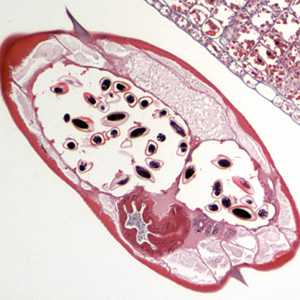
Figure A
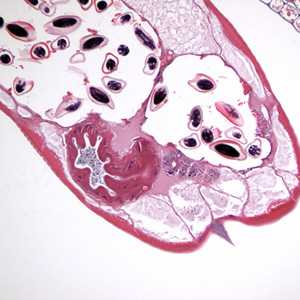
Figure B
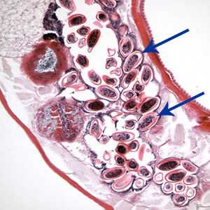
Figure C
Case Answer
This was a case of enterobiasis caused by Enterobius vermicularis (pinworm). Diagnostic features were:
- the presence of prominent lateral alae.
- the presence of oval eggs flattened on one side.
- the size of the eggs, which was consistent with pinworm eggs.
More on: Enterobiasis (Pinworm)
This case was kindly contributed by Dr. R Worthington at Sheboygan Memorial Hospital.
Images presented in the monthly case studies are from specimens submitted for diagnosis or archiving. On rare occasions, clinical histories given may be partly fictitious.
DPDx is an education resource designed for health professionals and laboratory scientists. For an overview including prevention and control visit www.cdc.gov/parasites/.
- Page last reviewed: August 24, 2016
- Page last updated: August 24, 2016
- Content source:
- Global Health – Division of Parasitic Diseases and Malaria
- Notice: Linking to a non-federal site does not constitute an endorsement by HHS, CDC or any of its employees of the sponsors or the information and products presented on the site.
- Maintained By: