


Case #202 - April, 2007
 ShareCompartir
A patient was seen at a hospital in Rwanda with headache, fever, and chills. A thin blood smear was made, stained with Giemsa, and examined. Figures A-F show what was seen on the smear at 1000× magnification. What is your diagnosis? Based on what criteria?
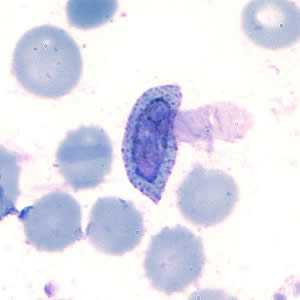
Figure A
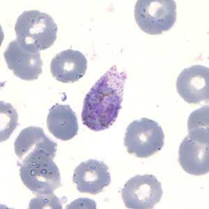
Figure B
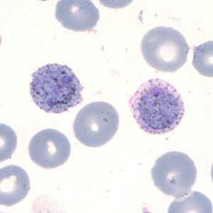
Figure C
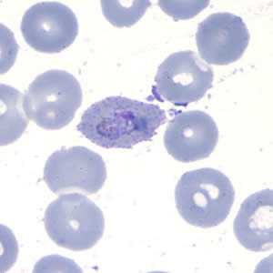
Figure D

Figure E
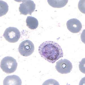
Figure F
Case Answer
This was a case of malaria caused by Plasmodium ovale. Many of the images depicted a classic presentation of P. ovale morphology; diagnostic features included:
- enlarged infected RBCs (1 1/4 to 1 1/2×).
- the presence of Schϋffner’s dots in the infected RBCs.
- elongated and fimbriated RBCs.
- the presence of multiple stages of Plasmodium, including:
- compact trophozoites with sturdy cytoplasm (Figure D)
- round to oval gametocytes that almost filled the RBCs (Figures A, C, and F)
- immature schizonts (Figures B and E). Immature schizonts are often not helpful for making a diagnosis because the merozoites are not fully developed and therefore cannot be counted. The size of the RBC can still be taken into consideration.
More on: Malaria
Case study images were captured from reference material donated by Mme. Severina Munyeshyaka of the University Hospital in Butare, Rwanda.
Images presented in the monthly case studies are from specimens submitted for diagnosis or archiving. On rare occasions, clinical histories given may be partly fictitious.
DPDx is an education resource designed for health professionals and laboratory scientists. For an overview including prevention and control visit www.cdc.gov/parasites/.
- Page last reviewed: August 24, 2016
- Page last updated: August 24, 2016
- Content source:
- Global Health – Division of Parasitic Diseases and Malaria
- Notice: Linking to a non-federal site does not constitute an endorsement by HHS, CDC or any of its employees of the sponsors or the information and products presented on the site.
- Maintained By: