


Case #197 - February, 2007
 ShareCompartir
The Division of Parasitic Diseases’ reference diagnostic laboratory received a specimen from a state health department for confirmatory review. The only patient information given was that the patient had traveled to Nigeria. Figures A-C show what was seen on a stained thin blood smear. What is your diagnosis? Based on what criteria?
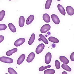
Figure A
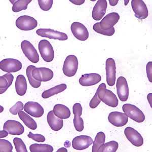
Figure B
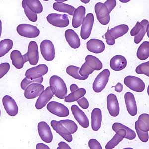
Figure C
Case Answer
This was a case of malaria caused by Plasmodium falciparum. Diagnostic features were:
- thin, delicate rings; some had double chromatin dots.
- an appliqué form (Figure B).
- the presence of only ring-stage trophozoites.
- infected red blood cells (RBCs) which did not appear to be enlarged.
In Plasmodium ovale infections, infected RBCs may be enlarged, distorted, fimbriated, roughly ovoid, and stippled. In this case, most infected and uninfected RBCs were ovoid, but the infected cells were not enlarged. The patient likely has hereditary elliptocytosis, a blood disorder in which a large proportion of RBCs are elliptical instead of bi-concave. This condition is caused by mutations in a number of different genes encoding cytoskeletal proteins of RBCs.
More on: Malaria
Images presented in the monthly case studies are from specimens submitted for diagnosis or archiving. On rare occasions, clinical histories given may be partly fictitious.
DPDx is an education resource designed for health professionals and laboratory scientists. For an overview including prevention and control visit www.cdc.gov/parasites/.
- Page last reviewed: August 24, 2016
- Page last updated: August 24, 2016
- Content source:
- Global Health – Division of Parasitic Diseases and Malaria
- Notice: Linking to a non-federal site does not constitute an endorsement by HHS, CDC or any of its employees of the sponsors or the information and products presented on the site.
- Maintained By: