


Case #196 - January, 2007
 ShareCompartir
A missionary traveled to various countries in sub-Saharan Africa for one year. He went to his physician complaining of abdominal pain and occasional diarrhea which he experienced since his return to the U.S. He reported he swam in various lakes while he was abroad. An ova and parasite (O & P) stool examination revealed objects, measuring 135 µm, like the one shown in Figures A and B (200×). The images show the same area of the slide but two separate focal planes. What is your diagnosis? Based on what criteria?
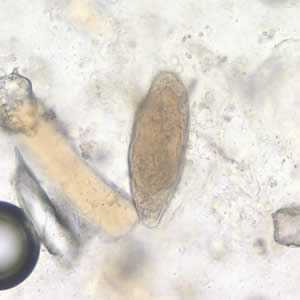
Figure A
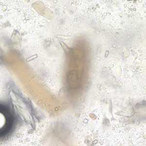
Figure B
Case Answer
This was a case of schistosomiasis caused by Schistosoma mansoni. Diagnostic features were:
- a miracidium is visible in the egg (Figure A, bracket).
- an unoperculated egg within the size range for S. mansoni (118-180 µm in length).
- presence of the typically prominent lateral spine, which was visible in a different focal plane (Figure B). Suspicious objects should be observed in different focal planes or key diagnostic features could be overlooked.
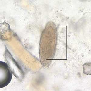
Figure A

Figure B
More on: Schistosomiasis
Images presented in the monthly case studies are from specimens submitted for diagnosis or archiving. On rare occasions, clinical histories given may be partly fictitious.
DPDx is an education resource designed for health professionals and laboratory scientists. For an overview including prevention and control visit www.cdc.gov/parasites/.
- Page last reviewed: August 24, 2016
- Page last updated: August 24, 2016
- Content source:
- Global Health – Division of Parasitic Diseases and Malaria
- Notice: Linking to a non-federal site does not constitute an endorsement by HHS, CDC or any of its employees of the sponsors or the information and products presented on the site.
- Maintained By: