


Case #190 - October, 2006
 ShareCompartir
The DPDx Team received a telediagnosis request asking for confirmation of an identification made by the Wisconsin State Laboratory of Hygiene. The patient had a travel history to India. Figures A and B show objects seen on the thick blood smears stained with Wright-Giemsa. Figures C-G were taken from the thin smears. What is your diagnosis? Based on what criteria?

Figure A
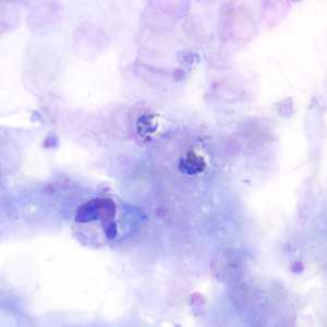
Figure B
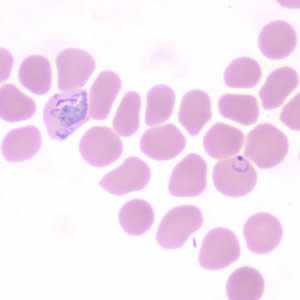
Figure C
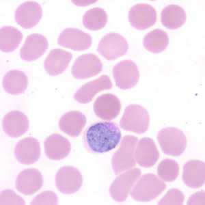
Figure D

Figure E
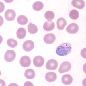
Figure F
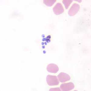
Figure G
Case Answer
This was a case of malaria caused by Plasmodium vivax. Diagnostic features were:
- sturdy rings (see Figures C and E).
- an amoeboid trophozoite (Figure C).
- a large gametocyte (Figure D) that almost fills the entire red blood cell. The infected cell is enlarged 1 1/2×.
- free merozoites following the rupture of a schizont (Figure G).
Depending on type of stain used and pH of the stain, Schüffner’s dots (stippling) may not always be demonstrated. For information on staining blood smears, please click here.
More on: Malaria
The images in this case were kindly contributed by the Wisconsin State Laboratory of Hygiene.
Images presented in the monthly case studies are from specimens submitted for diagnosis or archiving. On rare occasions, clinical histories given may be partly fictitious.
DPDx is an education resource designed for health professionals and laboratory scientists. For an overview including prevention and control visit www.cdc.gov/parasites/.
- Page last reviewed: August 24, 2016
- Page last updated: August 24, 2016
- Content source:
- Global Health – Division of Parasitic Diseases and Malaria
- Notice: Linking to a non-federal site does not constitute an endorsement by HHS, CDC or any of its employees of the sponsors or the information and products presented on the site.
- Maintained By: