


Case #189 - October, 2006
 ShareCompartir
A 4-year-old boy’s parents discovered a worm that they thought had been spit out by their child. They submitted the worm to their physician, along with the information that the boy had some exposure to raw seafood and that the family had a dog. The worm was eventually sent to a state public health laboratory and then CDC for identification. The worm was approximately 2 cm in length (see Figure A). The worm was cleared with lacto-phenol but defining morphologic features could not be seen. The posterior end was difficult to examine due to the cuticle being “rolled up” (see Figure B, 40× magnification). The anterior end was broken off (see Figure C, 40× magnification). A small cross-sectional slice of the worm was obtained with a scalpel and placed on a microscope slide with the cut side facing up. A small amount of lacto-phenol was placed around the section and a coverslip “floated” gently onto it, with additional solution along the edges of the coverslip to make an effective seal. Figure D, taken at 100× magnification shows what was observed in the section. What is your diagnosis? Based on what criteria?
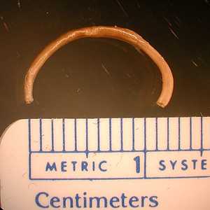
Figure A
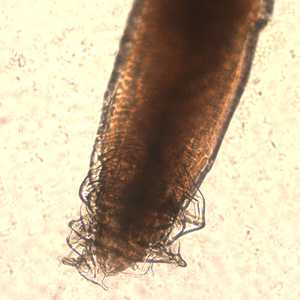
Figure B
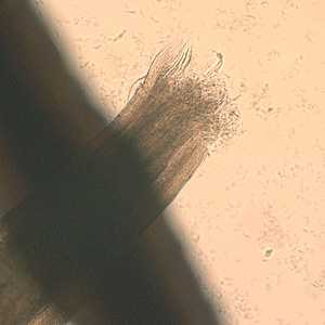
Figure C
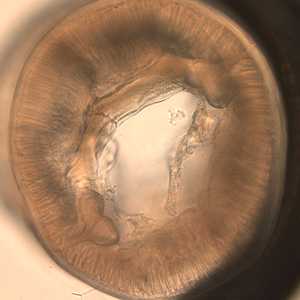
Figure D
Case Answer
This was a case of anisakiasis, caused by a nematode in the genus Pseudoterranova or Contracaecum. Diagnostic features were:
- the size range, which was consistent with L3 and L4 anisakid worms that have been reported as being coughed up.
- the large number of tall muscle cells, which is compatible with ascarid worms, including anisakids.
- the large butterfly-shaped lateral chords, indicative of Pseudoterranova and Contracaecum (see Figure A below, black arrows). Anisakis has lateral cords that are more delicate and Y-shaped.
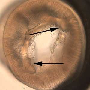
Figure A
Separation of Contracaecum and Pseudoterranova requires a detailed study of the digestive tract. Such structures were not intact enough to see clearly in this case.
More on: Anisakiasis
Images presented in the monthly case studies are from specimens submitted for diagnosis or archiving. On rare occasions, clinical histories given may be partly fictitious.
DPDx is an education resource designed for health professionals and laboratory scientists. For an overview including prevention and control visit www.cdc.gov/parasites/.
- Page last reviewed: August 24, 2016
- Page last updated: August 24, 2016
- Content source:
- Global Health – Division of Parasitic Diseases and Malaria
- Notice: Linking to a non-federal site does not constitute an endorsement by HHS, CDC or any of its employees of the sponsors or the information and products presented on the site.
- Maintained By: