


Case #188 - September, 2006
 ShareCompartir
A hospital submitted blood films to CDC’s reference laboratory for identification of the microfilariae that were seen on the films. The object in Figure A was seen on a Wright-Giemsa stained blood film and measured approximately 180 µm. The object in B was seen on a Giemsa stained blood film and measured approximately 240-250 µm. Both A and B were taken at 500× magnification. What is your diagnosis? Based on what criteria?
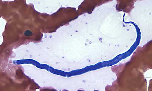
Figure A

Figure B
Case Answer
This was a case of loaisis caused by Loa loa. Diagnostic features were:
- a short cephalic space (red arrow, Figure A).
- the presence of nuclei to the tip of the tail (green arrow, Figure A).
- evidence of a sheath (black arrows, Figure A).
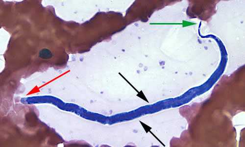
Figure A
More on: Loaisis
Images presented in the monthly case studies are from specimens submitted for diagnosis or archiving. On rare occasions, clinical histories given may be partly fictitious.
DPDx is an education resource designed for health professionals and laboratory scientists. For an overview including prevention and control visit www.cdc.gov/parasites/.
- Page last reviewed: August 24, 2016
- Page last updated: August 24, 2016
- Content source:
- Global Health – Division of Parasitic Diseases and Malaria
- Notice: Linking to a non-federal site does not constitute an endorsement by HHS, CDC or any of its employees of the sponsors or the information and products presented on the site.
- Maintained By: